
Deposition Date
2016-06-27
Release Date
2016-10-26
Last Version Date
2023-11-08
Entry Detail
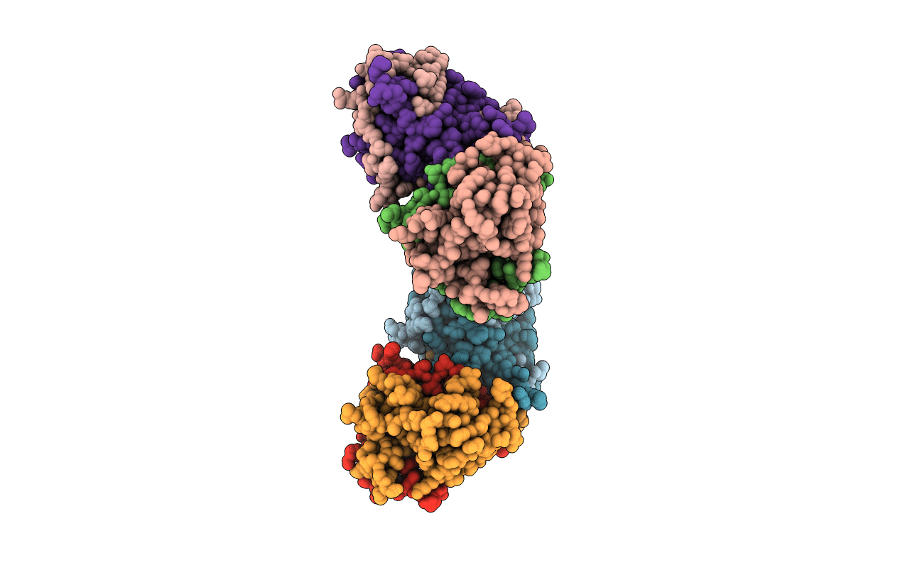
PDB ID:
5GJ4
Keywords:
Title:
Structure of NS2B-NS3 Protease from Zika Virus caught after self-cleavage
Biological Source:
Source Organism(s):
Zika virus (strain Mr 766) (Taxon ID: 64320)
Expression System(s):
Method Details:
Experimental Method:
Resolution:
1.84 Å
R-Value Free:
0.19
R-Value Work:
0.16
R-Value Observed:
0.18
Space Group:
P 31
