5GHL image
Deposition Date 2016-06-20
Release Date 2017-10-18
Last Version Date 2024-10-23
Entry Detail
PDB ID:
5GHL
Keywords:
Title:
Crystal structure Analysis of the starch-binding domain of glucoamylase from Aspergillus niger
Biological Source:
Source Organism:
Host Organism:
Method Details:
Experimental Method:
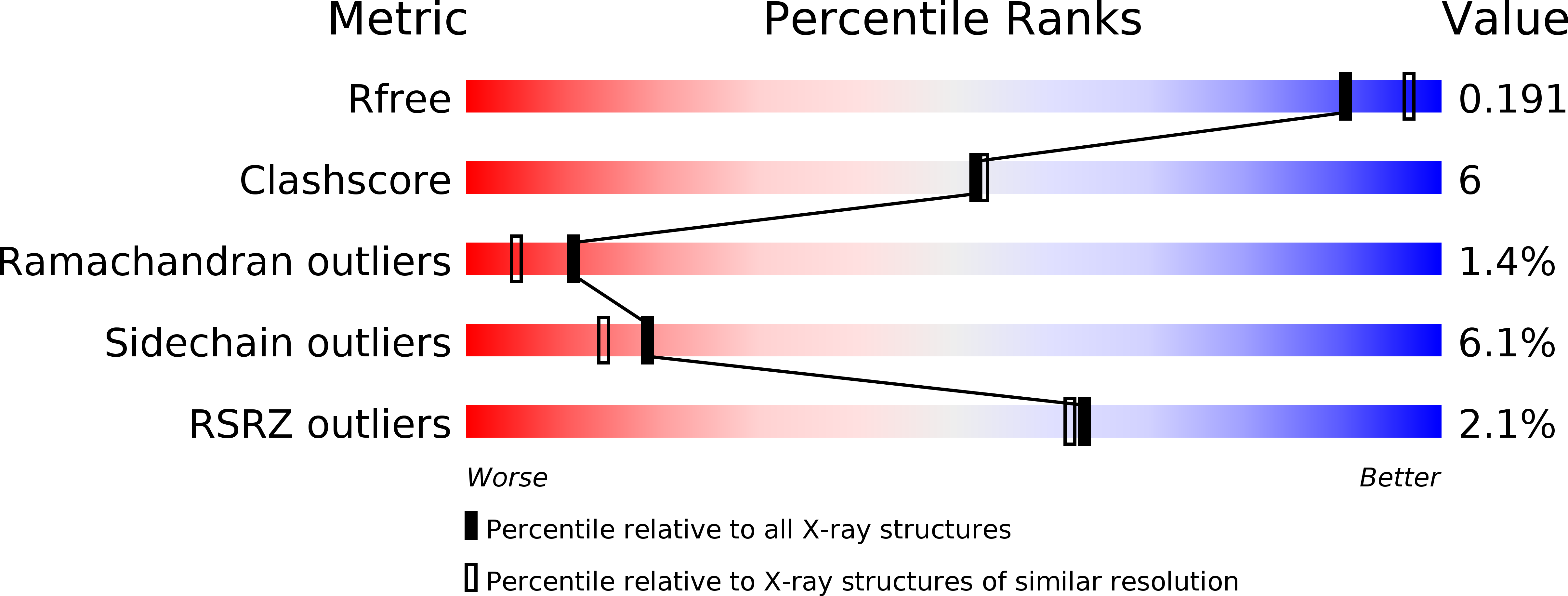
Resolution:
2.00 Å
R-Value Free:
0.22
R-Value Work:
0.17
R-Value Observed:
0.18
Space Group:
P 4
Macromolecular Entities
Structures with similar UniProt ID
Protein Blast
Polymer Type:polypeptide(L)
Molecule:Glucoamylase
Gene (Uniprot):GLAA
Chain IDs:A, B, C, D
Chain Length:108
Number of Molecules:4
Biological Source:Aspergillus niger
Primary Citation
Crystal structure of the starch-binding domain of glucoamylase from Aspergillus niger.
Acta Crystallogr.,Sect.F 73 550 554 (2017)
PMID: 28994402 DOI: 10.1107/S2053230X17012894

Abstact

Glucoamylases are widely used commercially to produce glucose syrup from starch. The starch-binding domain (SBD) of glucoamylase from Aspergillus niger is a small globular protein containing a disulfide bond. The structure of A. niger SBD has been determined by NMR, but the conformation surrounding the disulfide bond was unclear. Therefore, X-ray crystal structural analysis was used to attempt to clarify the conformation of this region. The SBD was purified from an Escherichia coli-based expression system and crystallized at 293 K. The initial phase was determined by the molecular-replacement method, and the asymmetric unit of the crystal contained four protomers, two of which were related by a noncrystallographic twofold axis. Finally, the structure was solved at 2.0 Å resolution. The SBD consisted of seven β-strands and eight loops, and the conformation surrounding the disulfide bond was determined from a clear electron-density map. Comparison of X-ray- and NMR-determined structures of the free SBD showed no significant difference in the conformation of each β-strand, but the conformations of the loops containing the disulfide bond and the L5 loop were different. In particular, the difference in the position of the Cα atom of Cys509 between the X-ray- and NMR-determined structures was 13.3 Å. In addition, the B factors of the amino-acid residues surrounding the disulfide bond are higher than those of other residues. Therefore, the conformation surrounding the disulfide bond is suggested to be highly flexible.

Legend

Protein

Chemical

Disease

Primary Citation of related structures