5G36 image
Deposition Date 2016-04-20
Release Date 2016-08-31
Last Version Date 2024-10-09
Entry Detail
PDB ID:
5G36
Title:
Yellow form of Halorhodopsin from Halobacterium salinarum in a new rhombohedral crystal form
Biological Source:
Source Organism:
Method Details:
Experimental Method:
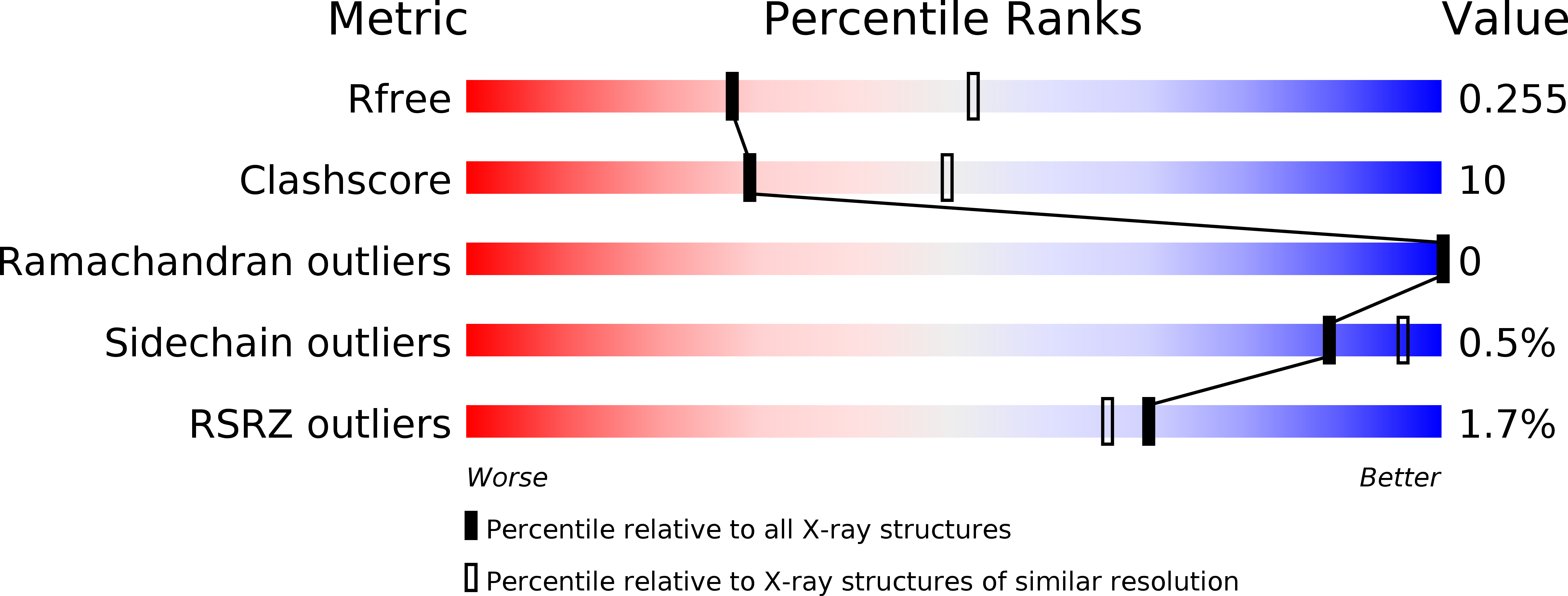
Resolution:
2.60 Å
R-Value Free:
0.25
R-Value Work:
0.19
R-Value Observed:
0.19
Space Group:
H 3 2
Macromolecular Entities
Structures with similar UniProt ID
Protein Blast
Polymer Type:polypeptide(L)
Molecule:HALORHODOPSIN
Gene (Uniprot):hop
Chain IDs:A
Chain Length:261
Number of Molecules:1
Biological Source:HALOBACTERIUM SALINARUM
Primary Citation
Crystal Structure of Halobacterium Salinarum Halorhodopsin with Partially Depopulated Primary Chloride Binding Site
Acta Crystallogr.,Sect.F 72 692 ? (2016)
PMID: 27599860 DOI: 10.1107/S2053230X16012796

Abstact

The transmembrane pump halorhodopsin in halophilic archaea translocates chloride ions from the extracellular to the cytoplasmic side upon illumination. In the ground state a tightly bound chloride ion occupies the primary chloride-binding site (CBS I) close to the protonated Schiff base that links the retinal chromophore to the protein. The light-triggered trans-cis isomerization of retinal causes structural changes in the protein associated with movement of the chloride ion. In reverse, chemical depletion of CBS I in Natronomonas pharaonis halorhodopsin (NpHR) through deprotonation of the Schiff base results in conformational changes of the protein: a state thought to mimic late stages of the photocycle. Here, crystals of Halobacterium salinarum halorhodopsin (HsHR) were soaked at high pH to provoke deprotonation of the Schiff base and loss of chloride. The crystals changed colour from purple to yellow and the occupancy of CBS I was reduced from 1 to about 0.5. In contrast to NpHR, this chloride depletion did not cause substantial conformational changes in the protein. Nevertheless, two observations indicate that chloride depletion could eventually result in structural changes similar to those found in NpHR. Firstly, the partially chloride-depleted form of HsHR has increased normalized B factors in the region of helix C that is close to CBS I and changes its conformation in NpHR. Secondly, prolonged soaking of HsHR crystals at high pH resulted in loss of diffraction. In conclusion, the conformation of the chloride-free protein may not be compatible with this crystal form of HsHR despite a packing arrangement that hardly restrains helices E and F that presumably move during ion transport.

Legend

Protein

Chemical

Disease

Primary Citation of related structures
Feedback Form
Name
Email
Institute
Feedback