Deposition Date
2016-02-05
Release Date
2016-06-29
Last Version Date
2024-01-10
Entry Detail
PDB ID:
5FVB
Keywords:
Title:
CRYSTAL STRUCTURE OF PHORMIDIUM C-PHYCOERYTHRIN AT PH 5.0
Biological Source:
Source Organism(s):
PHORMIDIUM RUBIDUM (Taxon ID: 865859)
Method Details:
Experimental Method:
Resolution:
1.93 Å
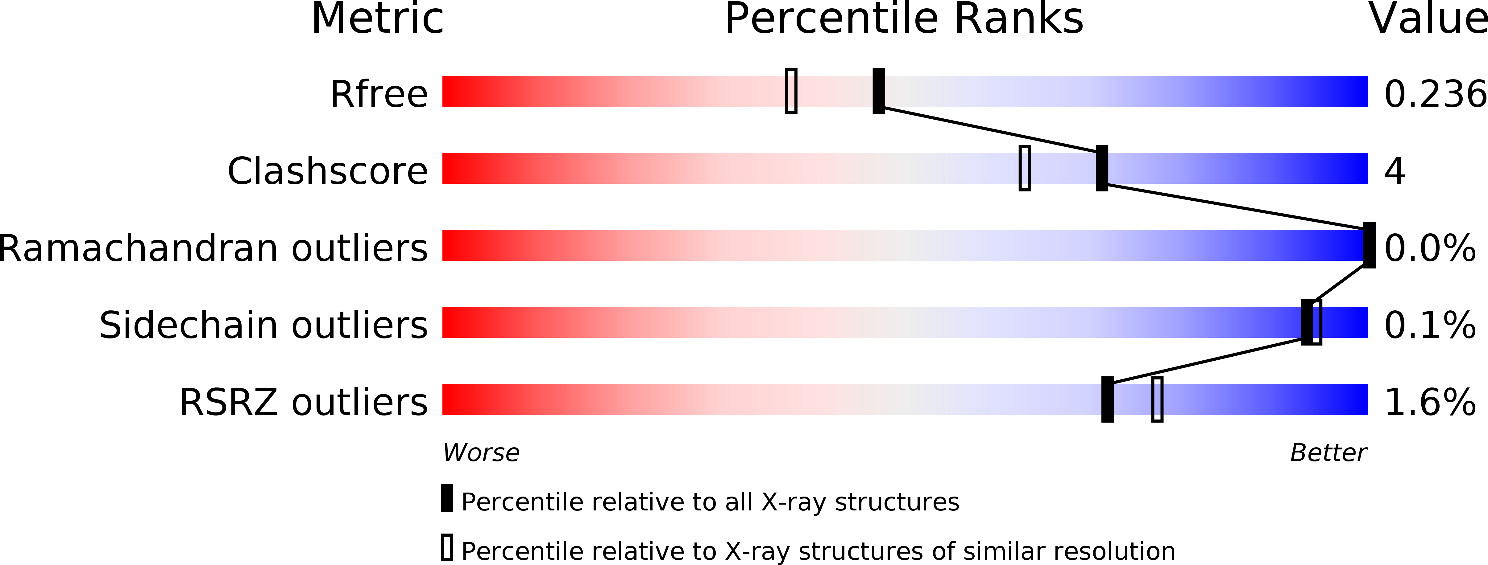
R-Value Free:
0.23
R-Value Work:
0.18
R-Value Observed:
0.18
Space Group:
P 1