5FTW image
Deposition Date 2016-01-18
Release Date 2016-10-05
Last Version Date 2024-01-10
Entry Detail
PDB ID:
5FTW
Keywords:
Title:
Crystal structure of glutamate O-methyltransferase in complex with S- adenosyl-L-homocysteine (SAH) from Bacillus subtilis
Biological Source:
Source Organism:
Host Organism:
Method Details:
Experimental Method:
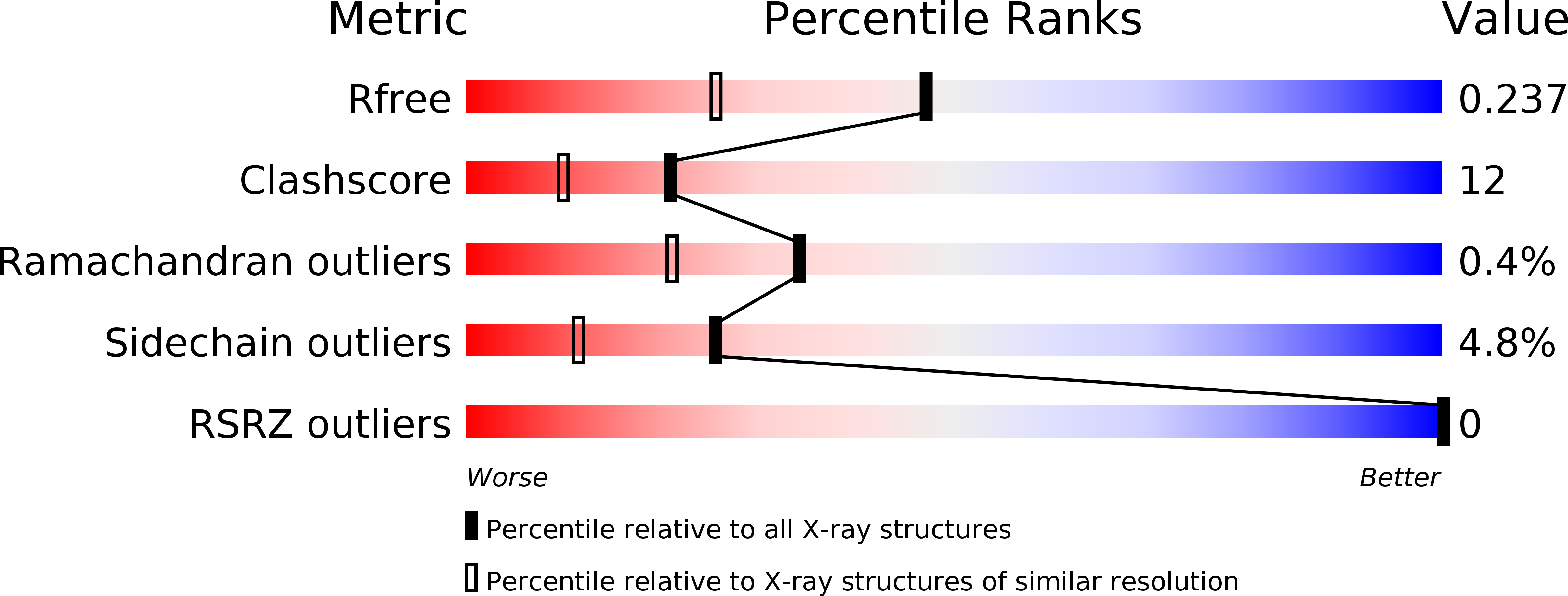
Resolution:
1.80 Å
R-Value Free:
0.23
R-Value Work:
0.17
R-Value Observed:
0.17
Space Group:
P 21 21 21
Macromolecular Entities
Structures with similar UniProt ID
Protein Blast
Polymer Type:polypeptide(L)
Molecule:CHEMOTAXIS PROTEIN METHYLTRANSFERASE
Gene (Uniprot):cheR
Chain IDs:A
Chain Length:256
Number of Molecules:1
Biological Source:BACILLUS SUBTILIS
Primary Citation
Crystal Structure of Pentapeptide-Independent Chemotaxis Receptor Methyltransferase (Cher) Reveals Idiosyncratic Structural Determinants for Receptor Recognition.
J.Struct.Biol. 196 364 ? (2016)
PMID: 27544050 DOI: 10.1016/J.JSB.2016.08.005

Abstact

Chemotactic methyltransferase, CheR catalyse methylation of specific glutamate residues in the cytoplasmic domain of methyl-accepting chemotactic protein receptors (MCPRs). The methylation of MCPRs is essential for the chemical sensing and chemotactic bacterial mobility towards favorable chemicals or away from unfavorable ones. In this study, crystal structure of B. subtilis CheR (BsCheR) in complex with S-adenosyl-l-homocysteine (SAH) has been determined to 1.8Å resolution. This is the first report of crystal structure belonging to the pentapeptide-independent CheR (PICheR) class. Till date, only one crystal structure of CheR from S. typhimurium (StCheR) belonging to pentapeptide-dependent CheR (PDCheR) class is available. Structural analysis of BsCheR reveals a helix-X-helix motif (HXH) with Asp53 as the linker residue in the N-terminal domain. The key structural features of the PDCheR β-subdomain involved in the formation of a tight complex with the pentapeptide binding motif in MCPRs were found to be absent in the structure of BsCheR. Additionally, isothermal titration calorimetry (ITC) experiments were performed to investigate S-adenosyl-(l)-methionine (SAM) binding affinity and KD was determined to be 0.32mM. The structure of BsCheR reveals that mostly residues of the large C-terminal domain contribute to SAH binding, with contributions of few residues from the linker region and the N-terminal domain. Structural investigations and sequence analysis carried out in this study provide critical insights into the distinct receptor recognition mechanism of the PDCheR and PICheR methyltransferase classes.

Legend

Protein

Chemical

Disease

Primary Citation of related structures