Deposition Date
2016-01-15
Release Date
2016-05-04
Last Version Date
2024-01-10
Entry Detail
PDB ID:
5FTU
Keywords:
Title:
Tetrameric complex of Latrophilin 3, Unc5D and FLRT2
Biological Source:
Source Organism(s):
RATTUS NORVEGICUS (Taxon ID: 10116)
MUS MUSCULUS (Taxon ID: 10090)
MUS MUSCULUS (Taxon ID: 10090)
Expression System(s):
Method Details:
Experimental Method:
Resolution:
6.01 Å
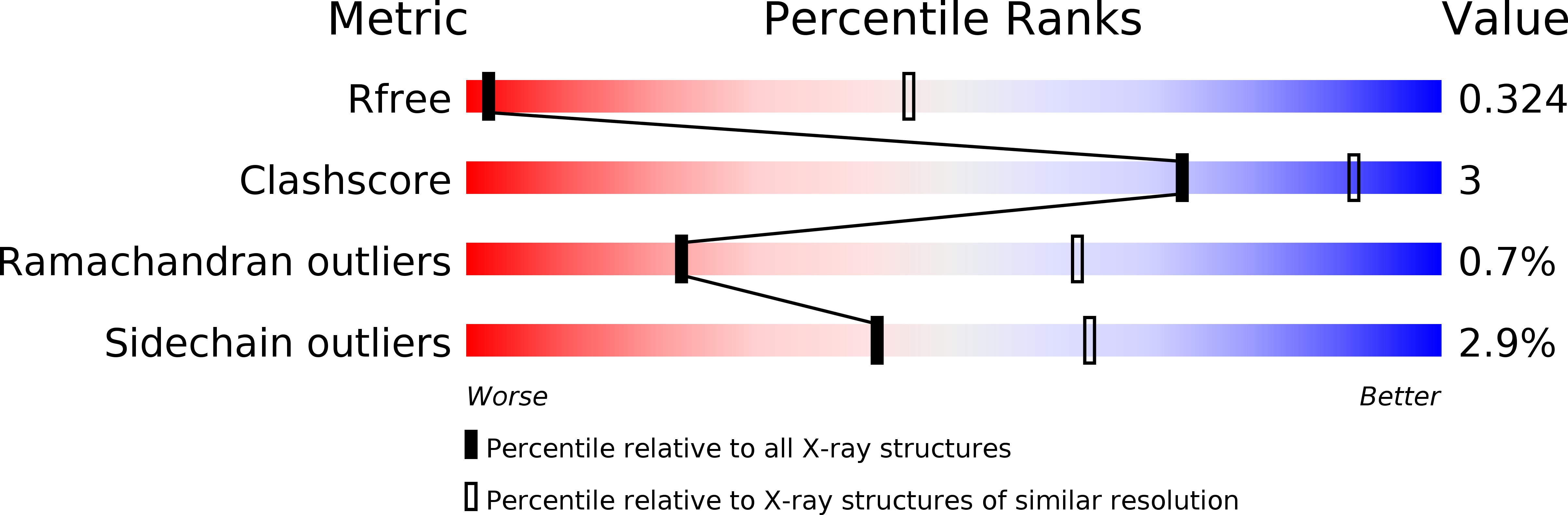
R-Value Free:
0.27
R-Value Work:
0.27
R-Value Observed:
0.27
Space Group:
I 41 2 2