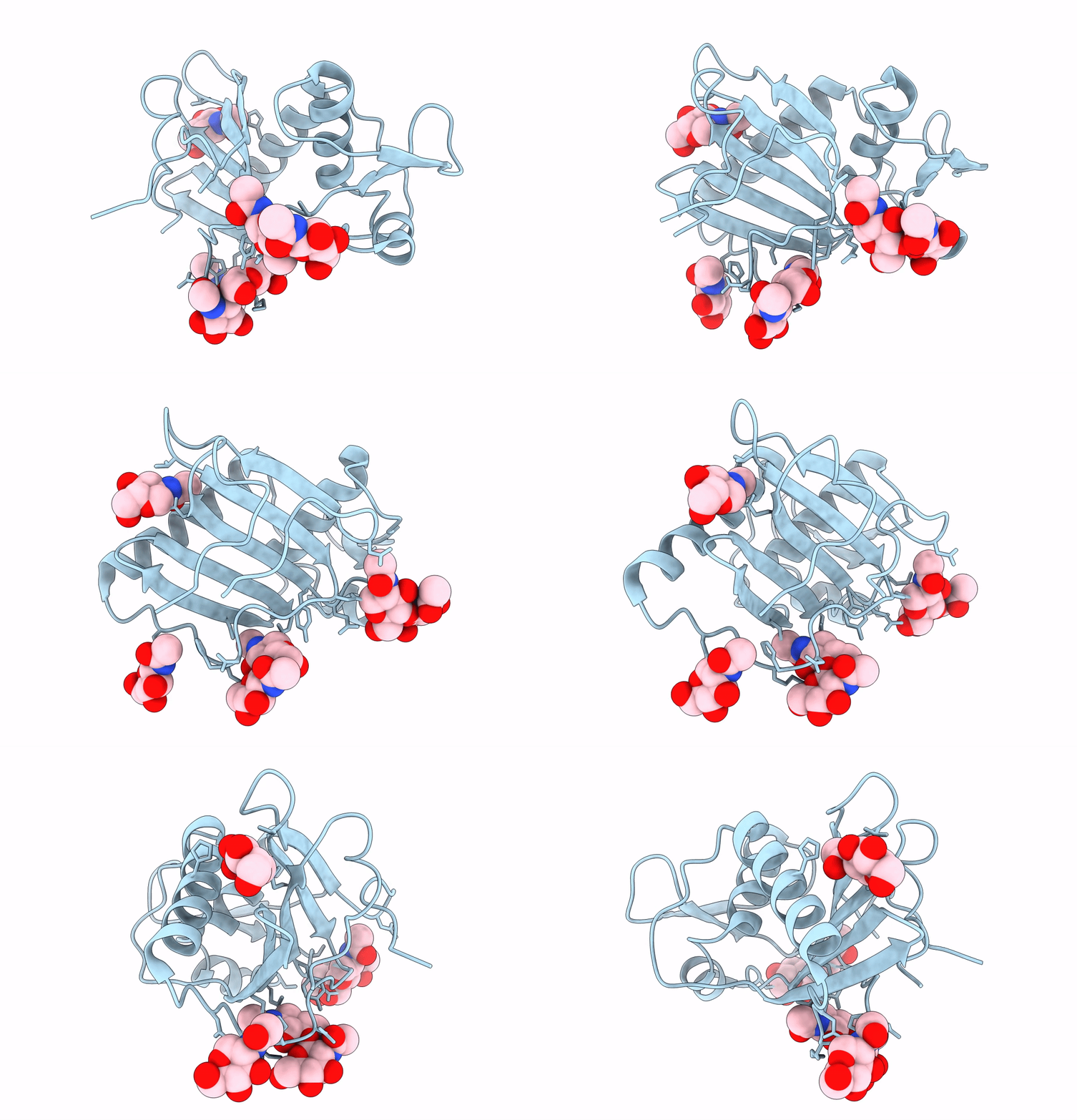
5FT2 image
Entry Detail
PDB ID:
5FT2
Keywords:
Title:
Sub-tomogram averaging of Lassa virus glycoprotein spike from virus- like particles at pH 5
Biological Source:
Source Organism:
Host Organism:
PDB Version:
Deposition Date:
2016-01-09
Release Date:
2016-03-02
Method Details:
Experimental Method:
Resolution:
16.40 Å
Aggregation State:
PARTICLE
Reconstruction Method:
TOMOGRAPHY
Macromolecular Entities
Polymer Type:polypeptide(L)
Description:PRE-GLYCOPROTEIN POLYPROTEIN GP COMPLEX
Chain IDs:A (auth: B)
Chain Length:172
Number of Molecules:1
Biological Source:LASSA VIRUS
Modified Residue
Compound ID Chain ID Parent Comp ID Details 2D Image
ASN A ASN GLYCOSYLATION SITE
Ligand Molecules
Primary Citation
Acidic Ph-Induced Conformations and Lamp1 Binding of the Lassa Virus Glycoprotein Spike.
Plos Pathog. 12 5418 ? (2016)
PMID: 26849049 DOI: 10.1371/JOURNAL.PPAT.1005418

Abstact

Lassa virus is an enveloped, bi-segmented RNA virus and the most prevalent and fatal of all Old World arenaviruses. Virus entry into the host cell is mediated by a tripartite surface spike complex, which is composed of two viral glycoprotein subunits, GP1 and GP2, and the stable signal peptide. Of these, GP1 binds to cellular receptors and GP2 catalyzes fusion between the viral envelope and the host cell membrane during endocytosis. The molecular structure of the spike and conformational rearrangements induced by low pH, prior to fusion, remain poorly understood. Here, we analyzed the three-dimensional ultrastructure of Lassa virus using electron cryotomography. Sub-tomogram averaging yielded a structure of the glycoprotein spike at 14-Å resolution. The spikes are trimeric, cover the virion envelope, and connect to the underlying matrix. Structural changes to the spike, following acidification, support a viral entry mechanism dependent on binding to the lysosome-resident receptor LAMP1 and further dissociation of the membrane-distal GP1 subunits.

Legend

Protein

Chemical

Disease

Primary Citation of related structures