5FT1 image
Deposition Date 2016-01-08
Release Date 2016-08-03
Last Version Date 2024-01-10
Entry Detail
PDB ID:
5FT1
Title:
Crystal structure of gp37(Dip) from bacteriophage phiKZ bound to RNase E of Pseudomonas aeruginosa
Biological Source:
Source Organism:
Host Organism:
Method Details:
Experimental Method:
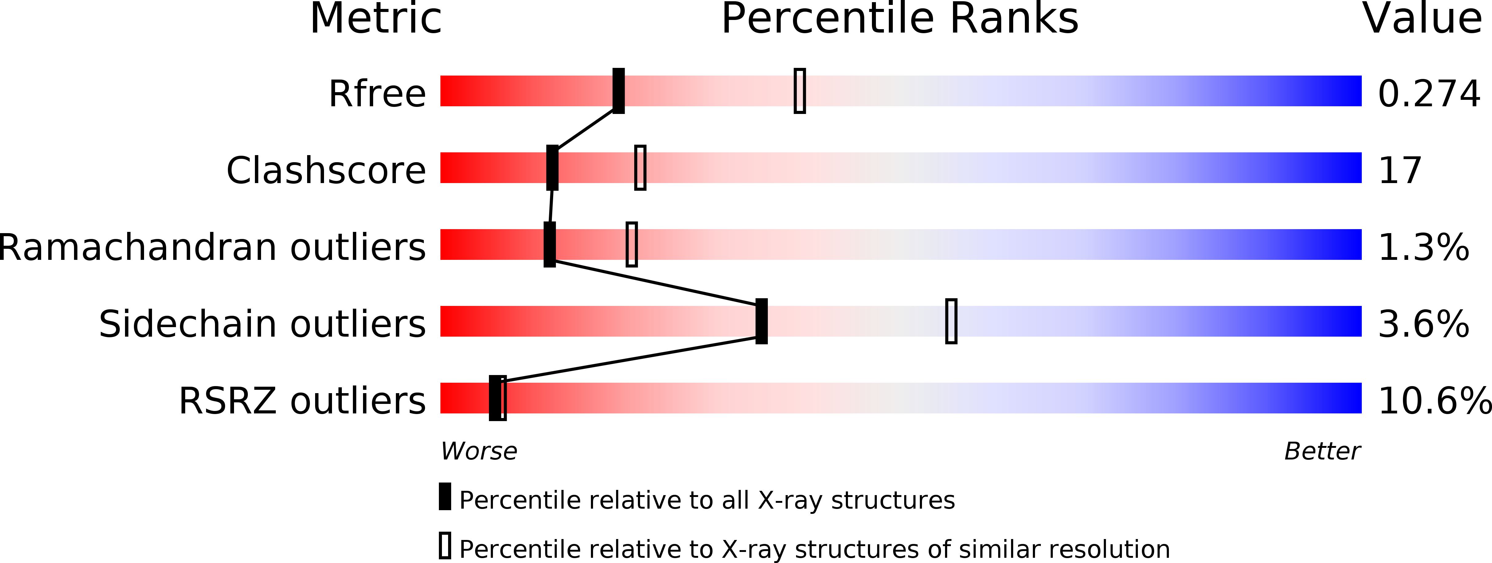
Resolution:
2.75 Å
R-Value Free:
0.29
R-Value Work:
0.26
R-Value Observed:
0.26
Space Group:
P 1
Macromolecular Entities
Polymer Type:polypeptide(L)
Molecule:GP37
Chain IDs:A, B, E, G, I, K
Chain Length:281
Number of Molecules:6
Biological Source:PSEUDOMONAS PHAGE PHIKZ
Polymer Type:polypeptide(L)
Molecule:RIBONUCLEASE E
Gene (Uniprot):rne
Chain IDs:C, D, F, H, J, L
Chain Length:20
Number of Molecules:6
Biological Source:PSEUDOMONAS AERUGINOSA
Primary Citation
Structural elucidation of a novel mechanism for the bacteriophage-based inhibition of the RNA degradosome.
Elife 5 ? ? (2016)
PMID: 27447594 DOI: 10.7554/eLife.16413

Abstact

In all domains of life, the catalysed degradation of RNA facilitates rapid adaptation to changing environmental conditions, while destruction of foreign RNA is an important mechanism to prevent host infection. We have identified a virus-encoded protein termed gp37/Dip, which directly binds and inhibits the RNA degradation machinery of its bacterial host. Encoded by giant phage фKZ, this protein associates with two RNA binding sites of the RNase E component of the Pseudomonas aeruginosa RNA degradosome, occluding them from substrates and resulting in effective inhibition of RNA degradation and processing. The 2.2 Å crystal structure reveals that this novel homo-dimeric protein has no identifiable structural homologues. Our biochemical data indicate that acidic patches on the convex outer surface bind RNase E. Through the activity of Dip, фKZ has evolved a unique mechanism to down regulate a key metabolic process of its host to allow accumulation of viral RNA in infected cells.

Legend

Protein

Chemical

Disease

Primary Citation of related structures