5FPQ image
Entry Detail
PDB ID:
5FPQ
Keywords:
Title:
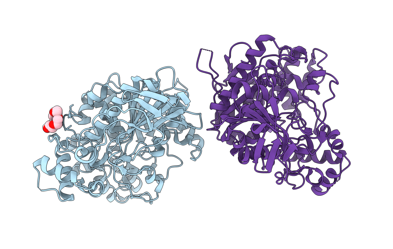
Structure of Homo sapiens acetylcholinesterase phosphonylated by sarin.
Biological Source:
Source Organism:
Host Organism:
PDB Version:
Deposition Date:
2015-12-02
Release Date:
2016-05-11
Method Details:
Experimental Method:
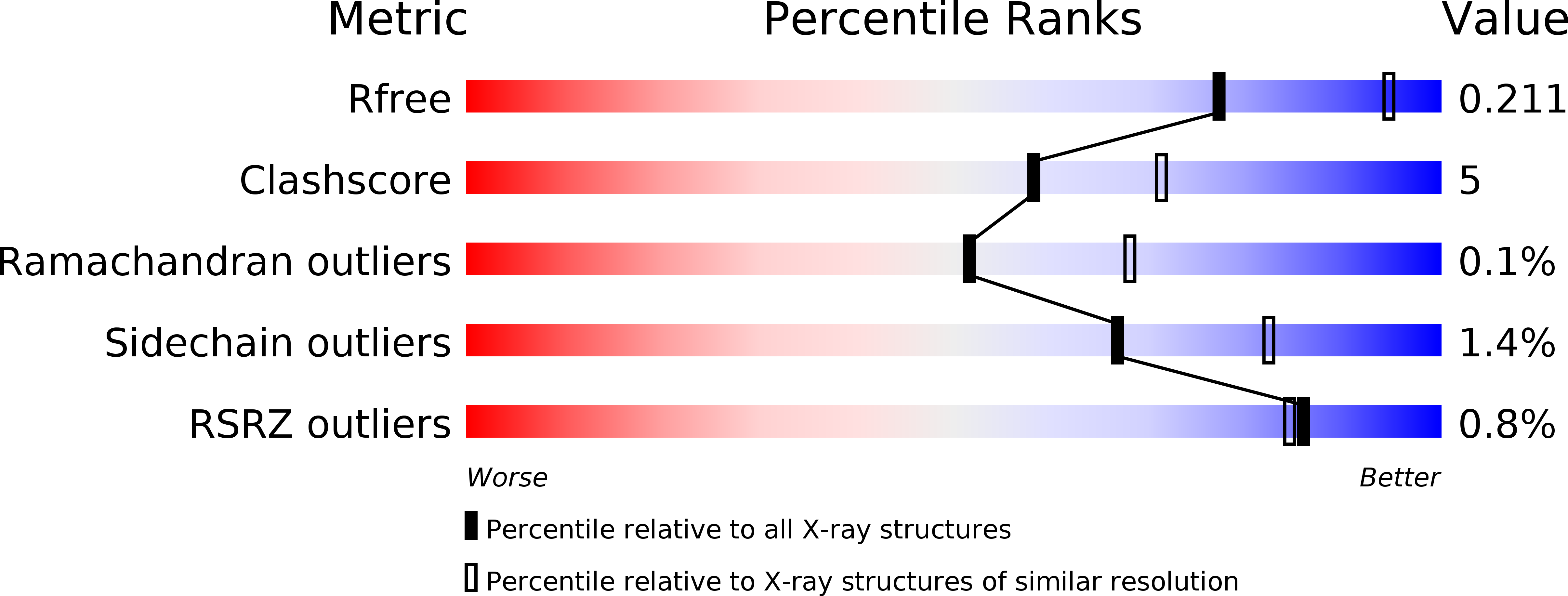
Resolution:
2.40 Å
R-Value Free:
0.21
R-Value Work:
0.17
R-Value Observed:
0.17
Space Group:
P 31 2 1
Macromolecular Entities
Polymer Type:polypeptide(L)
Description:ACETYLCHOLINESTERASE
Chain IDs:A, B
Chain Length:542
Number of Molecules:2
Biological Source:HOMO SAPIENS
Modified Residue
Compound ID Chain ID Parent Comp ID Details 2D Image
SGB A SER ?
Ligand Molecules
Primary Citation
Structure of a Prereaction Complex between the Nerve Agent Sarin, its Biological Target Acetylcholinesterase, and the Antidote Hi-6.
Proc.Natl.Acad.Sci.USA 113 5514 ? (2016)
PMID: 27140636 DOI: 10.1073/PNAS.1523362113

Abstact

Organophosphorus nerve agents interfere with cholinergic signaling by covalently binding to the active site of the enzyme acetylcholinesterase (AChE). This inhibition causes an accumulation of the neurotransmitter acetylcholine, potentially leading to overstimulation of the nervous system and death. Current treatments include the use of antidotes that promote the release of functional AChE by an unknown reactivation mechanism. We have used diffusion trap cryocrystallography and density functional theory (DFT) calculations to determine and analyze prereaction conformers of the nerve agent antidote HI-6 in complex with Mus musculus AChE covalently inhibited by the nerve agent sarin. These analyses reveal previously unknown conformations of the system and suggest that the cleavage of the covalent enzyme-sarin bond is preceded by a conformational change in the sarin adduct itself. Together with data from the reactivation kinetics, this alternate conformation suggests a key interaction between Glu202 and the O-isopropyl moiety of sarin. Moreover, solvent kinetic isotope effect experiments using deuterium oxide reveal that the reactivation mechanism features an isotope-sensitive step. These findings provide insights into the reactivation mechanism and provide a starting point for the development of improved antidotes. The work also illustrates how DFT calculations can guide the interpretation, analysis, and validation of crystallographic data for challenging reactive systems with complex conformational dynamics.

Legend

Protein

Chemical

Disease

Primary Citation of related structures