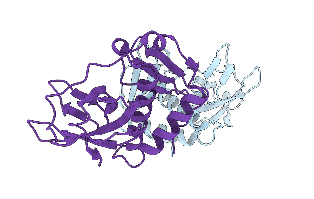
5FO3 image
Entry Detail
PDB ID:
5FO3
Keywords:
Title:
ZapC cell division regulator from E. coli
Biological Source:
Source Organism:
Host Organism:
PDB Version:
Deposition Date:
2015-11-18
Release Date:
2015-11-25
Method Details:
Experimental Method:
Resolution:
2.90 Å
R-Value Free:
0.24
R-Value Work:
0.21
R-Value Observed:
0.21
Space Group:
P 43 21 2
Macromolecular Entities
Polymer Type:polypeptide(L)
Description:CELL DIVISION PROTEIN ZAPC
Chain IDs:A, B
Chain Length:180
Number of Molecules:2
Biological Source:ESCHERICHIA COLI
Modified Residue
Compound ID Chain ID Parent Comp ID Details 2D Image
MSE A MET SELENOMETHIONINE
Ligand Molecules
Primary Citation
Crystal Structure of the Z-Ring Associated Cell Division Protein Zapc from Escherichia Coli.
FEBS Lett. 589 3822 ? (2015)
PMID: 26619764 DOI: 10.1016/J.FEBSLET.2015.11.030

Abstact

Bacterial cell division involves a contractile ring that organises downstream proteins at the division site and which contains the tubulin homologue FtsZ. ZapC has been discovered as a non-essential regulator of FtsZ. It localises to the septal ring and deletion of zapC leads to a mild phenotype, while overexpression inhibits cell division. Interference with cell division is facilitated by an interaction with FtsZ. Here, we present the 2.9 Å crystal structure of ZapC from Escherichia coli. ZapC forms a dimer and comprises two domains that belong to the Royal superfamily of which many members bind methylated arginines or lysines. ZapC contains an N-terminal chromo-like domain and a Tudor-like C-terminal domain. We show by ITC that ZapC binds the C-terminal tail of FtsZ.

Legend

Protein

Chemical

Disease

Primary Citation of related structures