Deposition Date
2015-11-13
Release Date
2016-07-13
Last Version Date
2024-11-06
Entry Detail
PDB ID:
5FNE
Keywords:
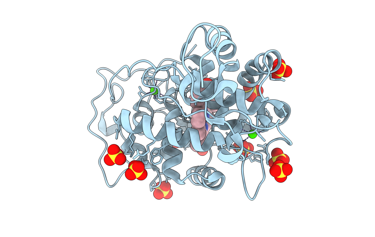
Title:
CRYSTAL STRUCTURE OF FUNGAL VERSATILE PEROXIDASE FROM PLEUROTUS ERYNGII TRIPLE MUTANT E37K, H39R & G330R
Biological Source:
Source Organism(s):
PLEUROTUS ERYNGII (Taxon ID: 5323)
Expression System(s):
Method Details:
Experimental Method:
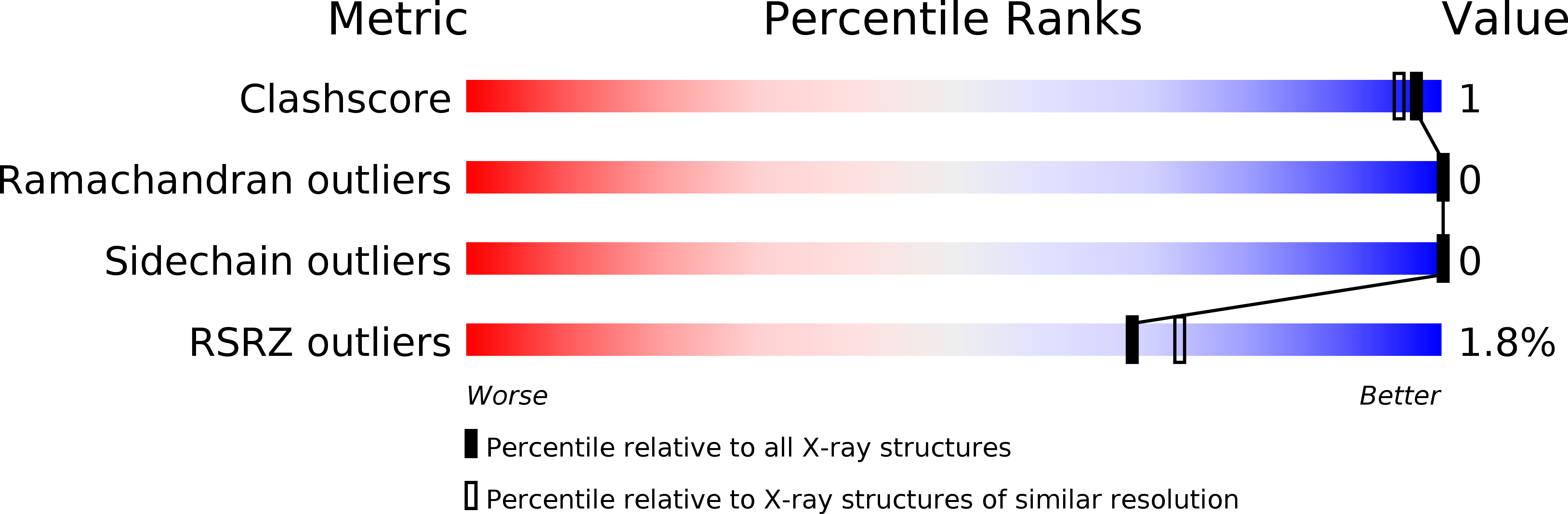
Resolution:
1.50 Å
R-Value Free:
0.22
R-Value Work:
0.19
R-Value Observed:
0.19
Space Group:
P 21 21 2