
Deposition Date
2015-10-22
Release Date
2016-06-29
Last Version Date
2024-01-10
Entry Detail
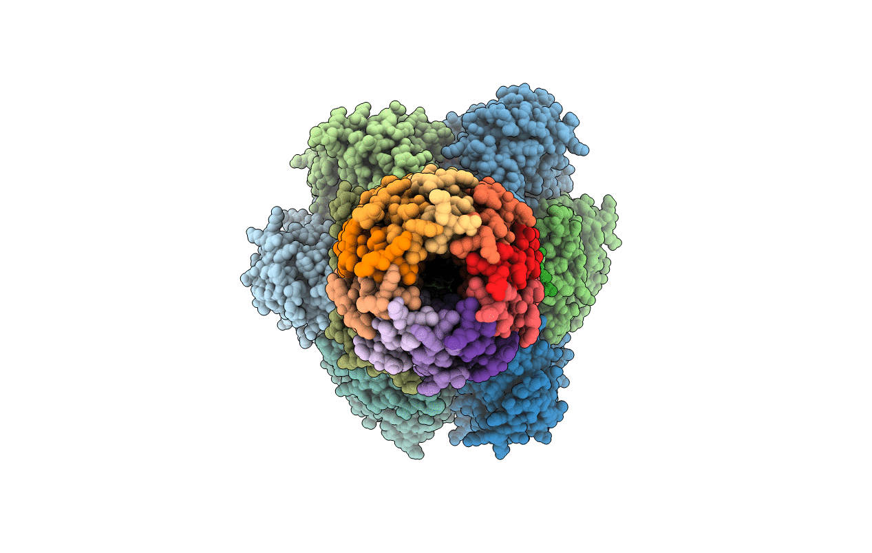
PDB ID:
5FL7
Keywords:
Title:
Structure of the F1c10 complex from Yarrowia lipolytica ATP synthase
Biological Source:
Source Organism(s):
YARROWIA LIPOLYTICA (Taxon ID: 4952)
Method Details:
Experimental Method:
Resolution:
3.50 Å
R-Value Free:
0.30
R-Value Work:
0.27
R-Value Observed:
0.27
Space Group:
P 21 21 2
