5FHY image
Deposition Date 2015-12-22
Release Date 2016-10-05
Last Version Date 2024-04-03
Entry Detail
PDB ID:
5FHY
Title:
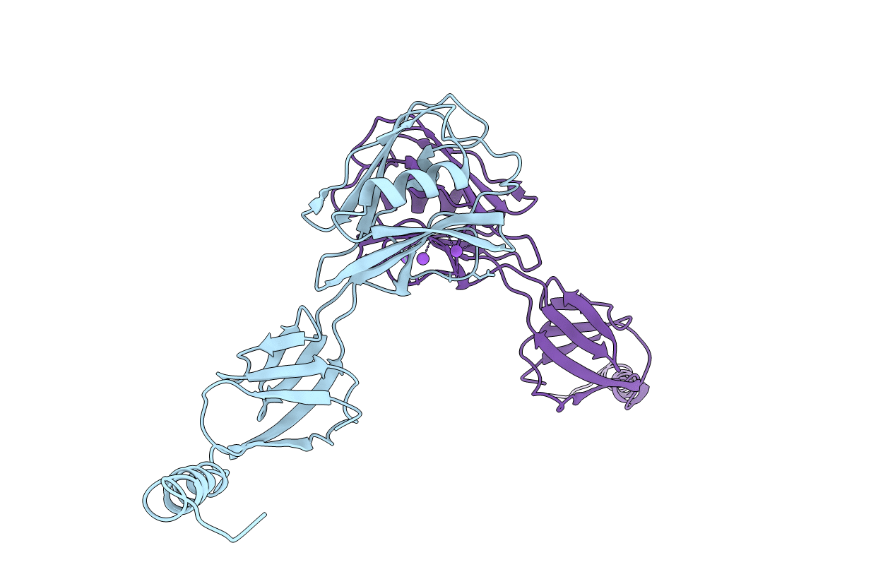
Crystal structure of FliD (HAP2) from Pseudomonas aeruginosa PAO1
Biological Source:
Method Details:
Experimental Method:
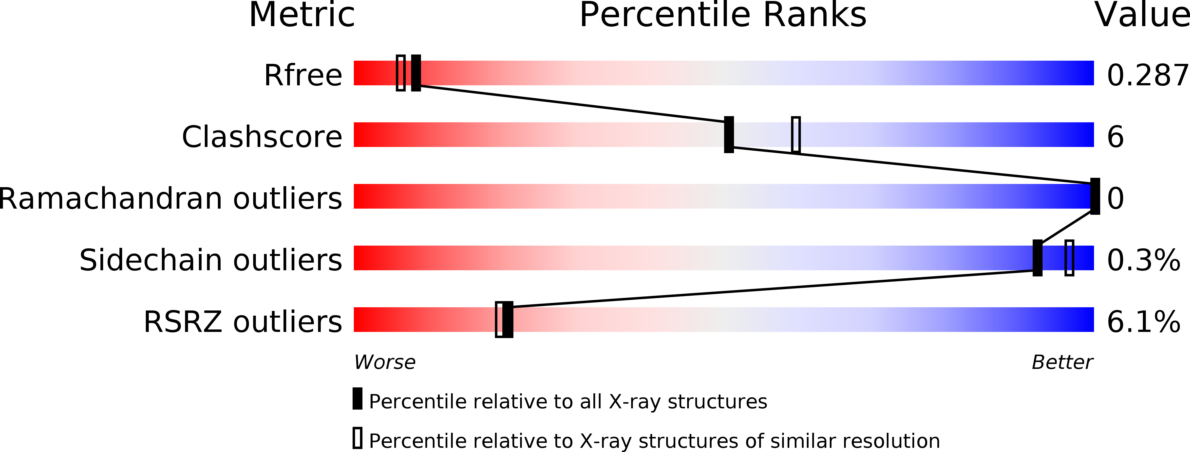
Resolution:
2.20 Å
R-Value Free:
0.28
R-Value Work:
0.24
R-Value Observed:
0.25
Space Group:
P 6
Macromolecular Entities
Structures with similar UniProt ID
Protein Blast
Polymer Type:polypeptide(L)
Molecule:B-type flagellar hook-associated protein 2
Gene (Uniprot):fliD
Chain IDs:A, B
Chain Length:346
Number of Molecules:2
Biological Source:Pseudomonas aeruginosa (strain ATCC 15692 / PAO1 / 1C / PRS 101 / LMG 12228)
Ligand Molecules
Primary Citation
Bacterial flagellar capping proteins adopt diverse oligomeric states.
Elife 5 ? ? (2016)
PMID: 27664419 DOI: 10.7554/eLife.18857

Abstact

Flagella are crucial for bacterial motility and pathogenesis. The flagellar capping protein (FliD) regulates filament assembly by chaperoning and sorting flagellin (FliC) proteins after they traverse the hollow filament and exit the growing flagellum tip. In the absence of FliD, flagella are not formed, resulting in impaired motility and infectivity. Here, we report the 2.2 Å resolution X-ray crystal structure of FliD from Pseudomonas aeruginosa, the first high-resolution structure of any FliD protein from any bacterium. Using this evidence in combination with a multitude of biophysical and functional analyses, we find that Pseudomonas FliD exhibits unexpected structural similarity to other flagellar proteins at the domain level, adopts a unique hexameric oligomeric state, and depends on flexible determinants for oligomerization. Considering that the flagellin filaments on which FliD oligomers are affixed vary in protofilament number between bacteria, our results suggest that FliD oligomer stoichiometries vary across bacteria to complement their filament assemblies.

Legend

Protein

Chemical

Disease

Primary Citation of related structures