Deposition Date
2015-12-22
Release Date
2017-01-11
Last Version Date
2024-01-10
Entry Detail
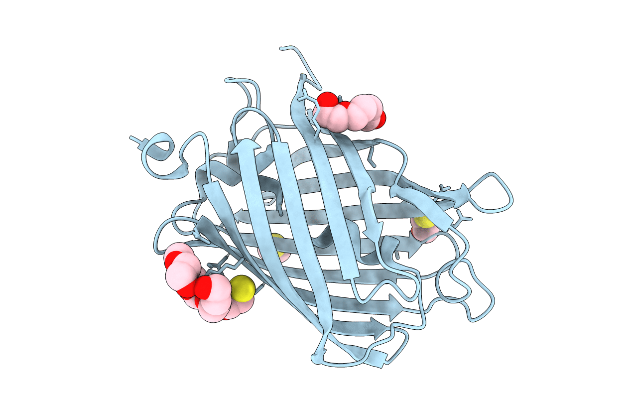
PDB ID:
5FHV
Keywords:
Title:
Crystal structure of mCherry after reaction with 2-mercaptoethanol
Biological Source:
Source Organism(s):
Discosoma sp. (Taxon ID: 86600)
Expression System(s):
Method Details:
Experimental Method:
Resolution:
1.55 Å
R-Value Free:
0.18
R-Value Work:
0.15
R-Value Observed:
0.15
Space Group:
P 1 21 1