5FEB image
Deposition Date 2015-12-16
Release Date 2016-03-09
Last Version Date 2024-10-09
Entry Detail
PDB ID:
5FEB
Title:
Crystal structure of the Voltage-gated Sodium Channel Beta 2 subunit extracellular domain
Biological Source:
Source Organism:
Homo sapiens (Taxon ID: 9606)
Host Organism:
Method Details:
Experimental Method:
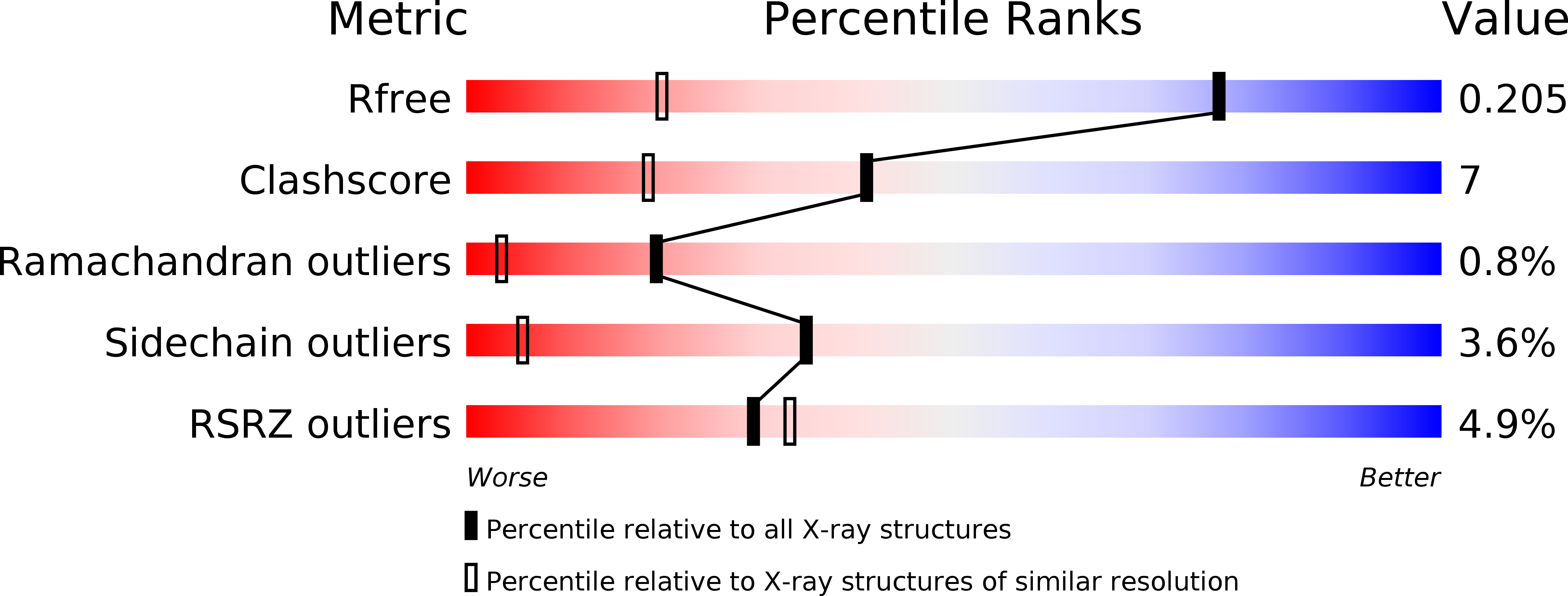
Resolution:
1.35 Å
R-Value Free:
0.20
R-Value Work:
0.17
R-Value Observed:
0.17
Space Group:
P 21 21 21
Macromolecular Entities
Structures with similar UniProt ID
Protein Blast
Polymer Type:polypeptide(L)
Molecule:Sodium channel subunit beta-2
Gene (Uniprot):SCN2B
Mutagens:C55A
Chain IDs:A
Chain Length:125
Number of Molecules:1
Biological Source:Homo sapiens
Ligand Molecules
Primary Citation
Binary architecture of the Nav1.2-beta 2 signaling complex.
Elife 5 ? ? (2016)
PMID: 26894959 DOI: 10.7554/eLife.10960

Abstact

To investigate the mechanisms by which β-subunits influence Nav channel function, we solved the crystal structure of the β2 extracellular domain at 1.35Å. We combined these data with known bacterial Nav channel structural insights and novel functional studies to determine the interactions of specific residues in β2 with Nav1.2. We identified a flexible loop formed by (72)Cys and (75)Cys, a unique feature among the four β-subunit isoforms. Moreover, we found that (55)Cys helps to determine the influence of β2 on Nav1.2 toxin susceptibility. Further mutagenesis combined with the use of spider toxins reveals that (55)Cys forms a disulfide bond with (910)Cys in the Nav1.2 domain II pore loop, thereby suggesting a 1:1 stoichiometry. Our results also provide clues as to which disulfide bonds are formed between adjacent Nav1.2 (912/918)Cys residues. The concepts emerging from this work will help to form a model reflecting the β-subunit location in a Nav channel complex.

Legend

Protein

Chemical

Disease

Primary Citation of related structures
Feedback Form
Name
Email
Institute
Feedback