5FCF image
Deposition Date 2015-12-15
Release Date 2016-12-07
Last Version Date 2024-03-20
Entry Detail
PDB ID:
5FCF
Keywords:
Title:
Crystal Structure of Xaa-Pro dipeptidase from Xanthomonas campestris, phosphate and Mn bound
Biological Source:
Method Details:
Experimental Method:
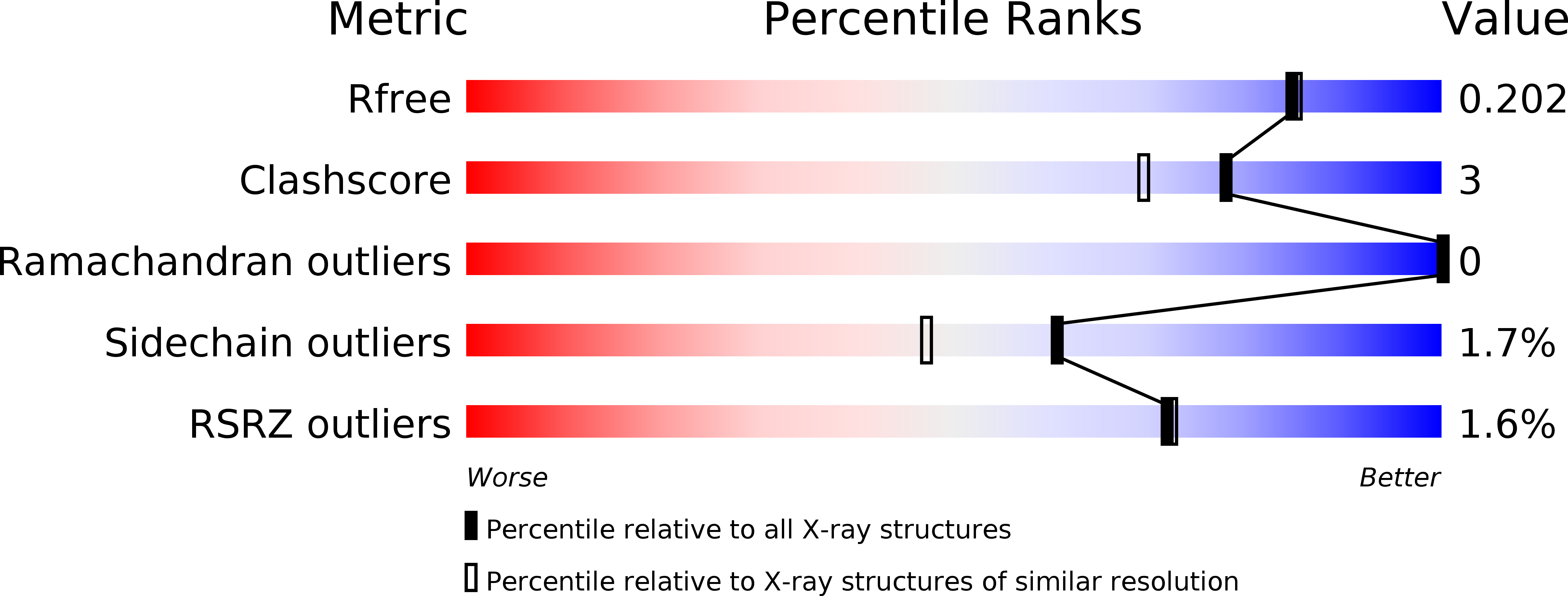
Resolution:
1.85 Å
R-Value Free:
0.19
R-Value Work:
0.15
R-Value Observed:
0.16
Space Group:
P 21 21 21
Macromolecular Entities
Structures with similar UniProt ID
Protein Blast
Polymer Type:polypeptide(L)
Molecule:Proline dipeptidase
Gene (Uniprot):pepQ
Chain IDs:A, B
Chain Length:399
Number of Molecules:2
Biological Source:Xanthomonas campestris pv. campestris str. ATCC 33913
Polymer Type:polypeptide(L)
Molecule:GLY-GLY-GLY
Chain IDs:C
Chain Length:3
Number of Molecules:1
Biological Source:Escherichia coli
Primary Citation
Crystal structure and biochemical investigations reveal novel mode of substrate selectivity and illuminate substrate inhibition and allostericity in a subfamily of Xaa-Pro dipeptidases.
Biochim. Biophys. Acta 1865 153 164 (2017)
PMID: 27816563 DOI: 10.1016/j.bbapap.2016.10.016

Abstact

Xaa-Pro dipeptidase (XPD) catalyzes hydrolysis of iminopeptide bond in dipeptides containing trans-proline as a second residue. XPDs are found in all living organisms and are believed to play an essential role in proline metabolism. Here, we report crystal structures and extensive enzymatic studies of XPD from Xanthomonas campestris (XPDxc), the first such comprehensive study of a bacterial XPD. We also report enzymatic activities of its ortholog from Mycobacterium tuberculosis (XPDmt). These enzymes are strictly dipeptidases with broad substrate specificities. They exhibit substrate inhibition and allostericity, as described earlier for XPD from Lactococcus lactis (XPDll). The structural, mutational and comparative data have revealed a novel mechanism of dipeptide selectivity and substrate binding in these enzymes. Moreover, we have identified conserved sequence motifs that distinguish these enzymes from other prolidases, thus defining a new subfamily. This study provides a suitable structural template for explaining unique properties of this XPDxc subfamily. In addition, we report unique structural features of XPDxc protein like an extended N-terminal tail region and absence of a conserved Tyr residue near the active site.

Legend

Protein

Chemical

Disease

Primary Citation of related structures