Deposition Date
2015-12-14
Release Date
2015-12-30
Last Version Date
2023-09-27
Entry Detail
PDB ID:
5FBU
Keywords:
Title:
Crystal structure of rifampin phosphotransferase RPH-Lm from Listeria monocytogenes in complex with rifampin-phosphate
Biological Source:
Source Organism(s):
Listeria monocytogenes serotype 4b str. F2365 (Taxon ID: 265669)
Expression System(s):
Method Details:
Experimental Method:
Resolution:
2.85 Å
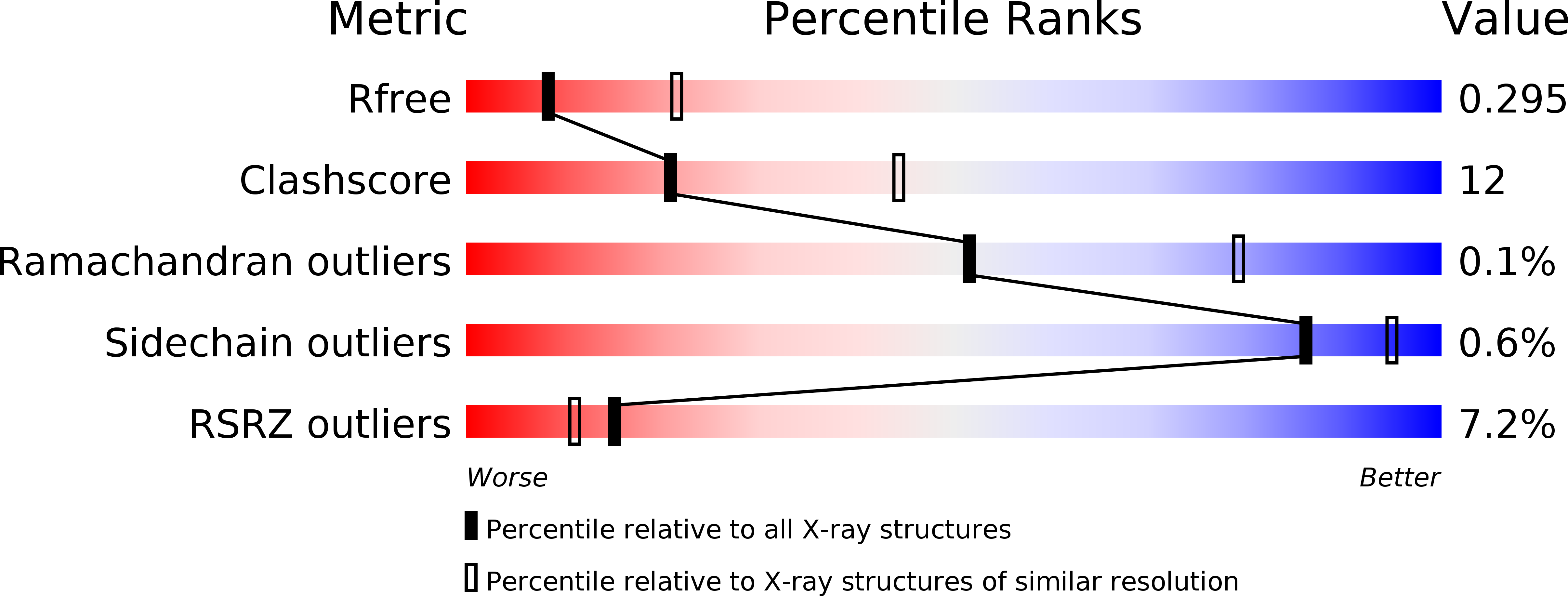
R-Value Free:
0.29
R-Value Work:
0.23
R-Value Observed:
0.24
Space Group:
P 65 2 2