5F7Z image
Entry Detail
PDB ID:
5F7Z
Keywords:
Title:
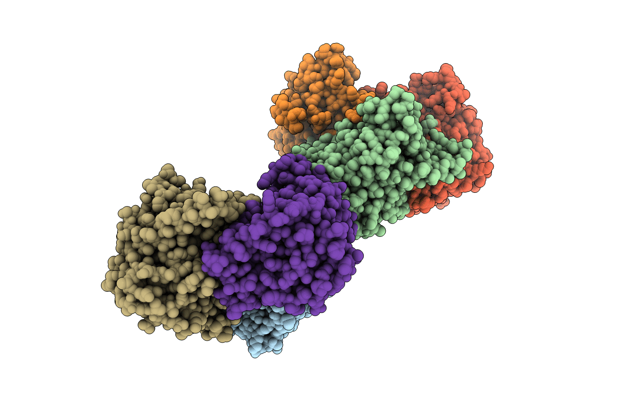
Crystal structure of Double Mutant S12T and N87T of Adenosine/Methylthioadenosine Phosphorylase from Schistosoma mansoni in APO Form
Biological Source:
Source Organism:
Host Organism:
PDB Version:
Deposition Date:
2015-12-08
Release Date:
2016-12-21
Method Details:
Experimental Method:
Resolution:
1.80 Å
R-Value Free:
0.23
R-Value Work:
0.21
R-Value Observed:
0.21
Space Group:
P 1 21 1
Macromolecular Entities
Polymer Type:polypeptide(L)
Description:Methylthioadenosine phosphorylase
Mutations:S12T,N87T
Chain IDs:A, B, C, D, E, F
Chain Length:320
Number of Molecules:6
Biological Source:Schistosoma mansoni
Ligand Molecules
Primary Citation
Crystal Structure of Schistosoma mansoni Adenosine Phosphorylase/5'-Methylthioadenosine Phosphorylase and Its Importance on Adenosine Salvage Pathway.
PLoS Negl Trop Dis 10 e0005178 e0005178 (2016)
PMID: 27935959 DOI: 10.1371/journal.pntd.0005178

Abstact

Schistosoma mansoni do not have de novo purine pathways and rely on purine salvage for their purine supply. It has been demonstrated that, unlike humans, the S. mansoni is able to produce adenine directly from adenosine, although the enzyme responsible for this activity was unknown. In the present work we show that S. mansoni 5´-deoxy-5´-methylthioadenosine phosphorylase (MTAP, E.C. 2.4.2.28) is capable of use adenosine as a substrate to the production of adenine. Through kinetics assays, we show that the Schistosoma mansoni MTAP (SmMTAP), unlike the mammalian MTAP, uses adenosine substrate with the same efficiency as MTA phosphorolysis, which suggests that this enzyme is part of the purine pathway salvage in S. mansoni and could be a promising target for anti-schistosoma therapies. Here, we present 13 SmMTAP structures from the wild type (WT), including three single and one double mutant, and generate a solid structural framework for structure description. These crystal structures of SmMTAP reveal that the active site contains three substitutions within and near the active site when compared to it mammalian counterpart, thus opening up the possibility of developing specific inhibitors to the parasite MTAP. The structural and kinetic data for 5 substrates reveal the structural basis for this interaction, providing substract for inteligent design of new compounds for block this enzyme activity.

Legend

Protein

Chemical

Disease

Primary Citation of related structures