
Deposition Date
2015-12-08
Release Date
2016-01-20
Last Version Date
2024-10-23
Entry Detail
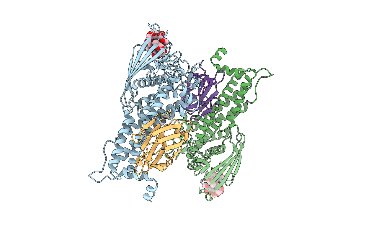
PDB ID:
5F7N
Keywords:
Title:
Blood group antigen binding adhesin BabA of Helicobacter pylori strain 17875 in complex with blood group A Lewis b pentasaccharide
Biological Source:
Source Organism(s):
Helicobacter pylori (Taxon ID: 210)
Vicugna pacos (Taxon ID: 30538)
Vicugna pacos (Taxon ID: 30538)
Expression System(s):
Method Details:
Experimental Method:
Resolution:
2.28 Å
R-Value Free:
0.19
R-Value Work:
0.17
R-Value Observed:
0.17
Space Group:
P 1 21 1
