5F19 image
Deposition Date 2015-11-30
Release Date 2016-03-16
Last Version Date 2024-11-20
Entry Detail
PDB ID:
5F19
Title:
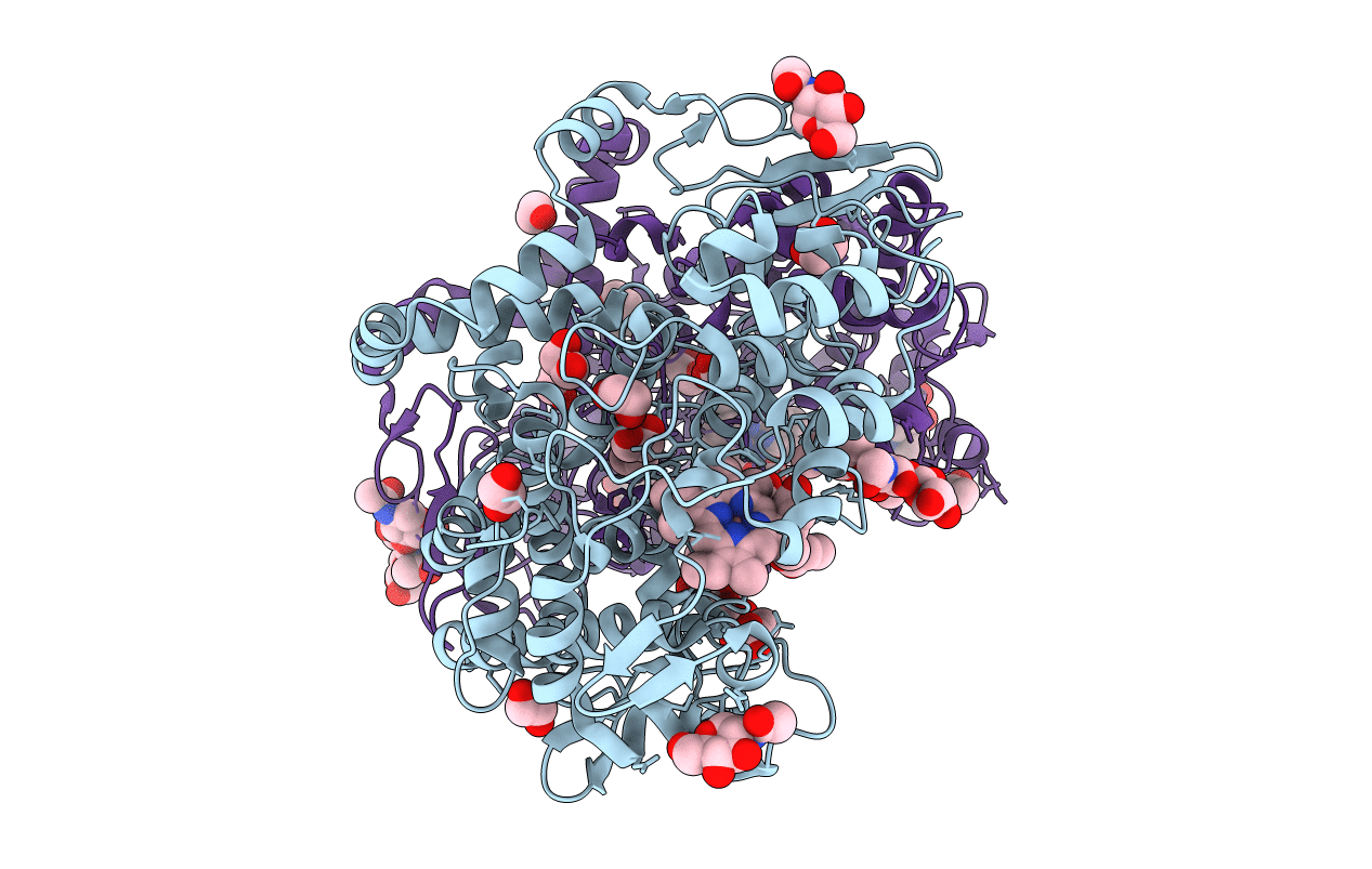
The Crystal Structure of Aspirin Acetylated Human Cyclooxygenase-2
Biological Source:
Source Organism:
Homo sapiens (Taxon ID: 9606)
Host Organism:
Method Details:
Experimental Method:
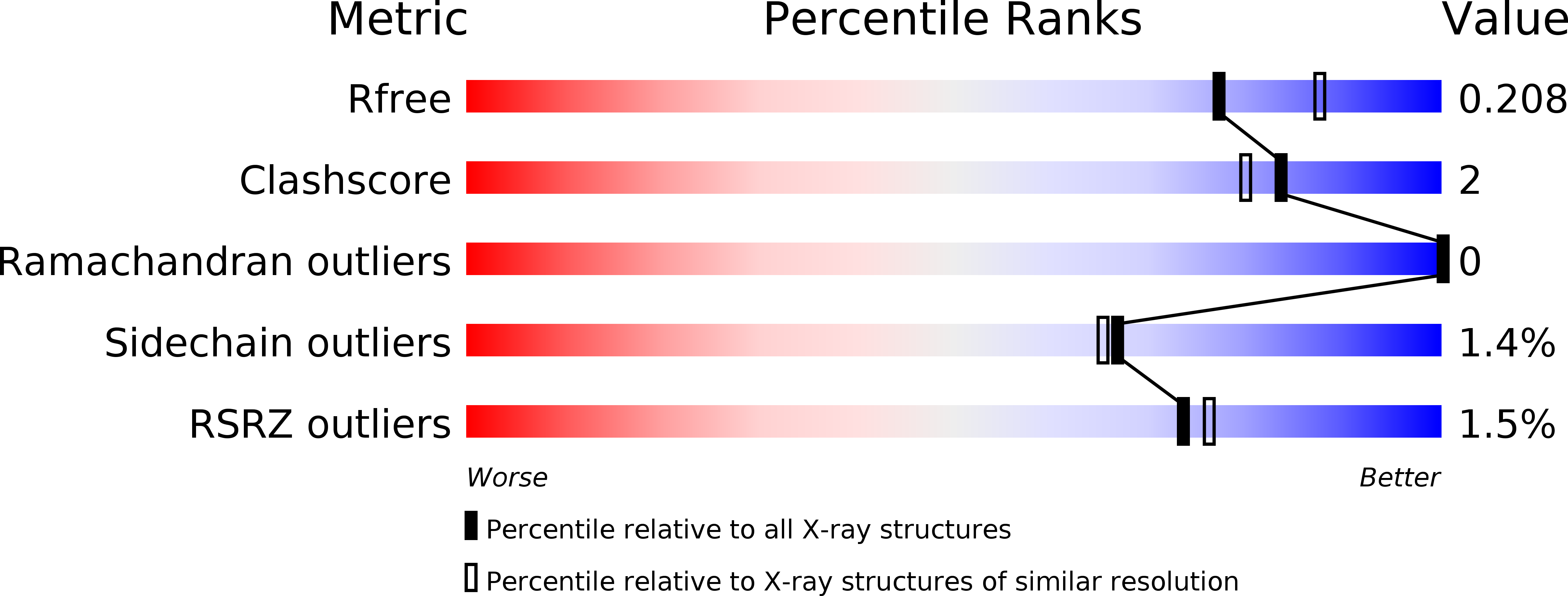
Resolution:
2.04 Å
R-Value Free:
0.20
R-Value Work:
0.16
R-Value Observed:
0.17
Space Group:
I 2 2 2
Macromolecular Entities
Polymer Type:polypeptide(L)
Molecule:Prostaglandin G/H synthase 2
Gene (Uniprot):PTGS2
Chain IDs:A, B
Chain Length:552
Number of Molecules:2
Biological Source:Homo sapiens
Modified Residue
Compound ID Chain ID Parent Comp ID Details 2D Image
OAS A SER modified residue
Primary Citation
Crystal Structure of Aspirin-Acetylated Human Cyclooxygenase-2: Insight into the Formation of Products with Reversed Stereochemistry.
Biochemistry 55 1226 1238 (2016)
PMID: 26859324 DOI: 10.1021/acs.biochem.5b01378

Abstact

Aspirin and other nonsteroidal anti-inflammatory drugs target the cyclooxygenase enzymes (COX-1 and COX-2) to block the formation of prostaglandins. Aspirin is unique in that it covalently modifies each enzyme by acetylating Ser-530 within the cyclooxygenase active site. Acetylation of COX-1 leads to complete loss of activity, while acetylation of COX-2 results in the generation of the monooxygenated product 15(R)-hydroxyeicosatetraenoic acid (15R-HETE). Ser-530 has also been shown to influence the stereochemistry for the addition of oxygen to the prostaglandin product. We determined the crystal structures of S530T murine (mu) COX-2, aspirin-acetylated human (hu) COX-2, and huCOX-2 in complex with salicylate to 1.9, 2.0, and 2.4 Å, respectively. The structures reveal that (1) the acetylated Ser-530 completely blocks access to the hydrophobic groove, (2) the observed binding pose of salicylate is reflective of the enzyme-inhibitor complex prior to acetylation, and (3) the observed Thr-530 rotamer in the S530T muCOX-2 crystal structure does not impede access to the hydrophobic groove. On the basis of these structural observations, along with functional analysis of the S530T/G533V double mutant, we propose a working hypothesis for the generation of 15R-HETE by aspirin-acetylated COX-2. We also observe differential acetylation of COX-2 purified in various detergent systems and nanodiscs, indicating that detergent and lipid binding within the membrane-binding domain of the enzyme alters the rate of the acetylation reaction in vitro.

Legend

Protein

Chemical

Disease

Primary Citation of related structures