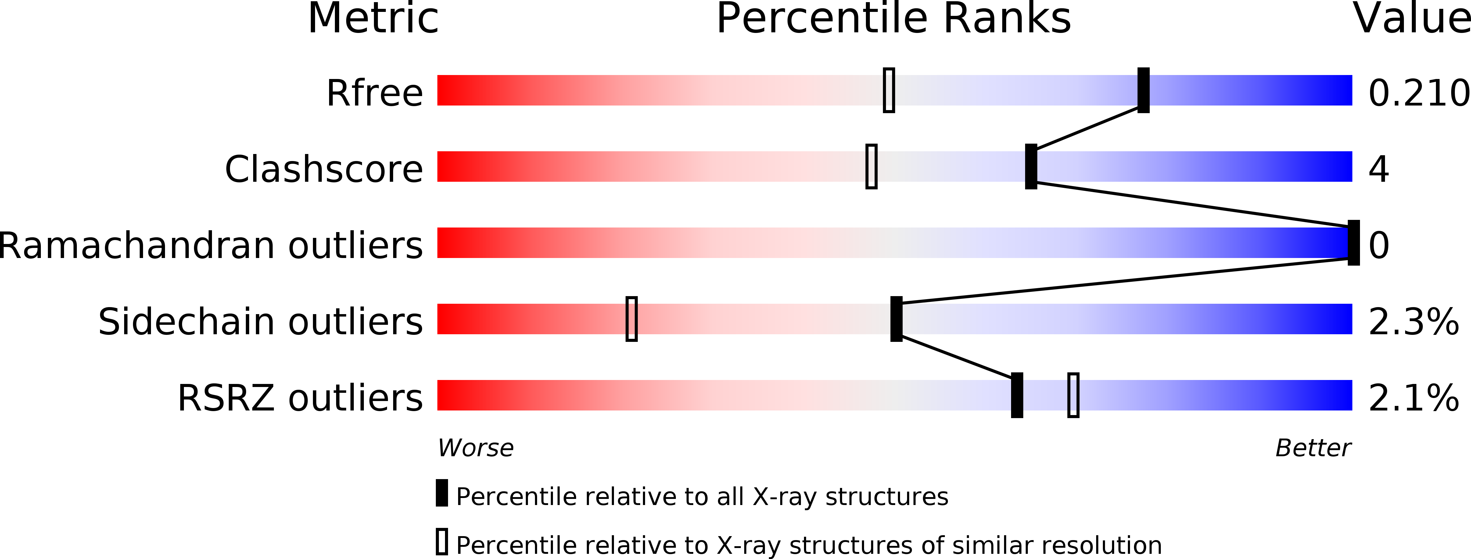
5EZU image
Deposition Date 2015-11-26
Release Date 2016-11-30
Last Version Date 2024-05-08
Entry Detail
PDB ID:
5EZU
Keywords:
Title:
Crystal structure of the N-terminal domain of vaccinia virus immunomodulator A46 in complex with myristic acid.
Biological Source:
Source Organism:
Method Details:
Experimental Method:
Resolution:
1.55 Å
R-Value Free:
0.24
R-Value Work:
0.19
R-Value Observed:
0.20
Space Group:
C 1 2 1
Macromolecular Entities
Polymer Type:polypeptide(L)
Molecule:Protein A46
Gene (Uniprot):OPG176
Chain IDs:A, B
Chain Length:89
Number of Molecules:2
Biological Source:Vaccinia virus (strain Western Reserve)
Ligand Molecules
Primary Citation
Vaccinia Virus Immunomodulator A46: A Lipid and Protein-Binding Scaffold for Sequestering Host TIR-Domain Proteins.
PLoS Pathog. 12 e1006079 e1006079 (2016)
PMID: 27973613 DOI: 10.1371/journal.ppat.1006079

Abstact

Vaccinia virus interferes with early events of the activation pathway of the transcriptional factor NF-kB by binding to numerous host TIR-domain containing adaptor proteins. We have previously determined the X-ray structure of the A46 C-terminal domain; however, the structure and function of the A46 N-terminal domain and its relationship to the C-terminal domain have remained unclear. Here, we biophysically characterize residues 1-83 of the N-terminal domain of A46 and present the X-ray structure at 1.55 Å. Crystallographic phases were obtained by a recently developed ab initio method entitled ARCIMBOLDO_BORGES that employs tertiary structure libraries extracted from the Protein Data Bank; data analysis revealed an all β-sheet structure. This is the first such structure solved by this method which should be applicable to any protein composed entirely of β-sheets. The A46(1-83) structure itself is a β-sandwich containing a co-purified molecule of myristic acid inside a hydrophobic pocket and represents a previously unknown lipid-binding fold. Mass spectrometry analysis confirmed the presence of long-chain fatty acids in both N-terminal and full-length A46; mutation of the hydrophobic pocket reduced the lipid content. Using a combination of high resolution X-ray structures of the N- and C-terminal domains and SAXS analysis of full-length protein A46(1-240), we present here a structural model of A46 in a tetrameric assembly. Integrating affinity measurements and structural data, we propose how A46 simultaneously interferes with several TIR-domain containing proteins to inhibit NF-κB activation and postulate that A46 employs a bipartite binding arrangement to sequester the host immune adaptors TRAM and MyD88.

Legend

Protein

Chemical

Disease

Primary Citation of related structures