5EZ2 image
Entry Detail
PDB ID:
5EZ2
Title:
Sandercyanin Fluorescent Protein (SFP)
Biological Source:
Source Organism:
PDB Version:
Deposition Date:
2015-11-26
Release Date:
2016-09-28
Method Details:
Experimental Method:
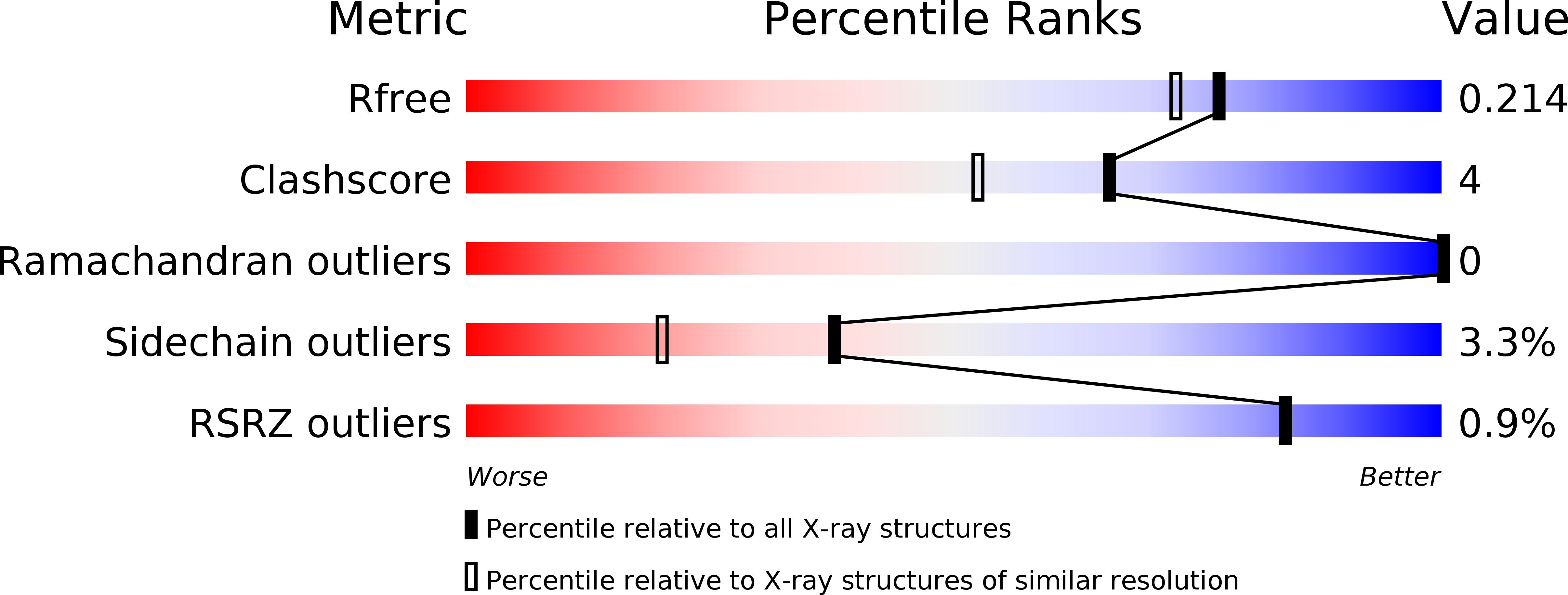
Resolution:
1.85 Å
R-Value Free:
0.21
R-Value Work:
0.19
R-Value Observed:
0.19
Space Group:
P 63 2 2
Macromolecular Entities
Polymer Type:polypeptide(L)
Description:Sandercyanin Fluorescent Protein
Chain IDs:A, B
Chain Length:183
Number of Molecules:2
Biological Source:Sander vitreus
Primary Citation
Blue protein with red fluorescence
Proc.Natl.Acad.Sci.USA 113 11513 11518 (2016)
PMID: 27688756 DOI: 10.1073/pnas.1525622113

Abstact

The walleye (Sander vitreus) is a golden yellow fish that inhabits the Northern American lakes. The recent sightings of the blue walleye and the correlation of its sighting to possible increased UV radiation have been proposed earlier. The underlying molecular basis of its adaptation to increased UV radiation is the presence of a protein (Sandercyanin)-ligand complex in the mucus of walleyes. Degradation of heme by UV radiation results in the formation of Biliverdin IXα (BLA), the chromophore bound to Sandercyanin. We show that Sandercyanin is a monomeric protein that forms stable homotetramers on addition of BLA to the protein. A structure of the Sandercyanin-BLA complex, purified from the fish mucus, reveals a glycosylated protein with a lipocalin fold. This protein-ligand complex absorbs light in the UV region (λmax of 375 nm) and upon excitation at this wavelength emits in the red region (λmax of 675 nm). Unlike all other known biliverdin-bound fluorescent proteins, the chromophore is noncovalently bound to the protein. We provide here a molecular rationale for the observed spectral properties of Sandercyanin.

Legend

Protein

Chemical

Disease

Primary Citation of related structures