Deposition Date
2015-11-13
Release Date
2016-06-08
Last Version Date
2023-09-27
Entry Detail
PDB ID:
5ER4
Keywords:
Title:
Crystal Structure of Calcium-loaded S100B bound to SC0025
Biological Source:
Source Organism(s):
Bos taurus (Taxon ID: 9913)
Expression System(s):
Method Details:
Experimental Method:
Resolution:
1.81 Å
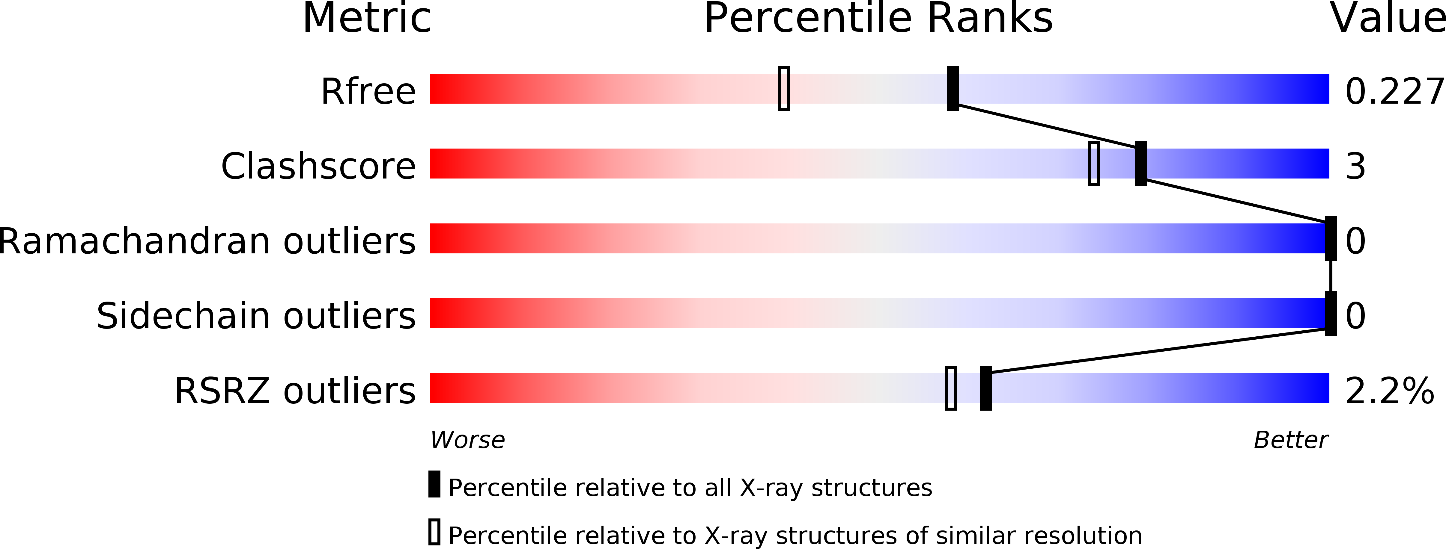
R-Value Free:
0.22
R-Value Work:
0.18
R-Value Observed:
0.18
Space Group:
C 2 2 21