5EQF image
Entry Detail
PDB ID:
5EQF
Keywords:
Title:
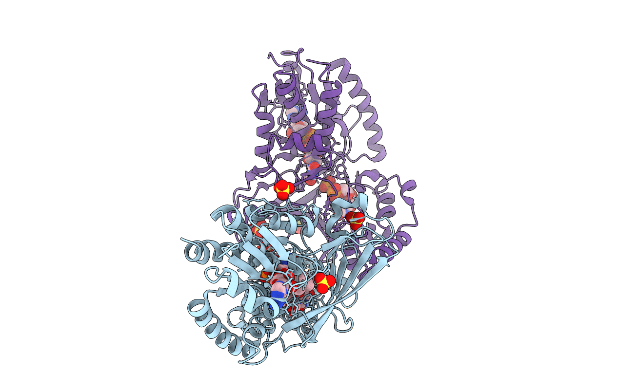
Crystal structure of oxidized UDP-galactopyranose mutase from Corynebacterium diphtheriae with UDP bound in closed form
Biological Source:
Host Organism:
PDB Version:
Deposition Date:
2015-11-12
Release Date:
2016-11-23
Method Details:
Experimental Method:
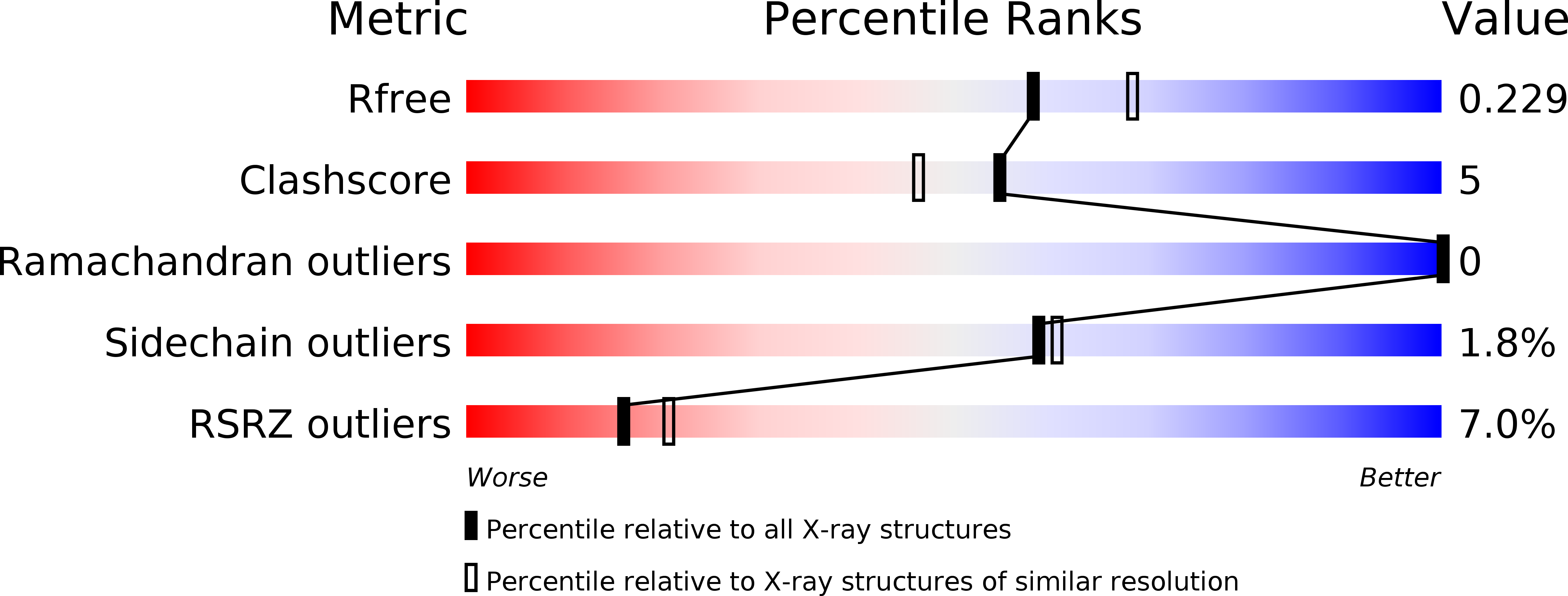
Resolution:
2.15 Å
R-Value Free:
0.22
R-Value Work:
0.17
R-Value Observed:
0.17
Space Group:
C 1 2 1
Macromolecular Entities
Polymer Type:polypeptide(L)
Description:UDP-galactopyranose mutase
Chain IDs:A, B
Chain Length:390
Number of Molecules:2
Biological Source:Corynebacterium diphtheriae
Primary Citation
Conformational Control of UDP-Galactopyranose Mutase Inhibition.
Biochemistry 56 3983 3992 (2017)
PMID: 28608671 DOI: 10.1021/acs.biochem.7b00189

Abstact

UDP-galactopyranose mutase (Glf or UGM) catalyzes the formation of uridine 5'-diphosphate-α-d-galactofuranose (UDP-Galf) from UDP-galactopyranose (UDP-Galp). The enzyme is required for the production of Galf-containing glycans. UGM is absent in mammals, but members of the Corynebacterineae suborder require UGM for cell envelope biosynthesis. The need for UGM in some pathogens has prompted the search for inhibitors that could serve as antibiotic leads. Optimizing inhibitor potency, however, has been challenging. The UGM from Klebsiella pneumoniae (KpUGM), which is not required for viability, is more effectively impeded by small-molecule inhibitors than are essential UGMs from species such as Mycobacterium tuberculosis or Corynebacterium diphtheriae. Why KpUGM is more susceptible to inhibition than other orthologs is not clear. One potential source of difference is UGM ortholog conformation. We previously determined a structure of CdUGM bound to a triazolothiadiazine inhibitor in the open form, but it was unclear whether the small-molecule inhibitor bound this form or to the closed form. By varying the terminal tag (CdUGM-His6 and GSG-CdUGM), we crystallized CdUGM to capture the enzyme in different conformations. These structures reveal a pocket in the active site that can be exploited to augment inhibitor affinity. Moreover, they suggest the inhibitor binds the open form of most prokaryotic UGMs but can bind the closed form of KpUGM. This model and the structures suggest strategies for optimizing inhibitor potency by exploiting UGM conformational flexibility.

Legend

Protein

Chemical

Disease

Primary Citation of related structures