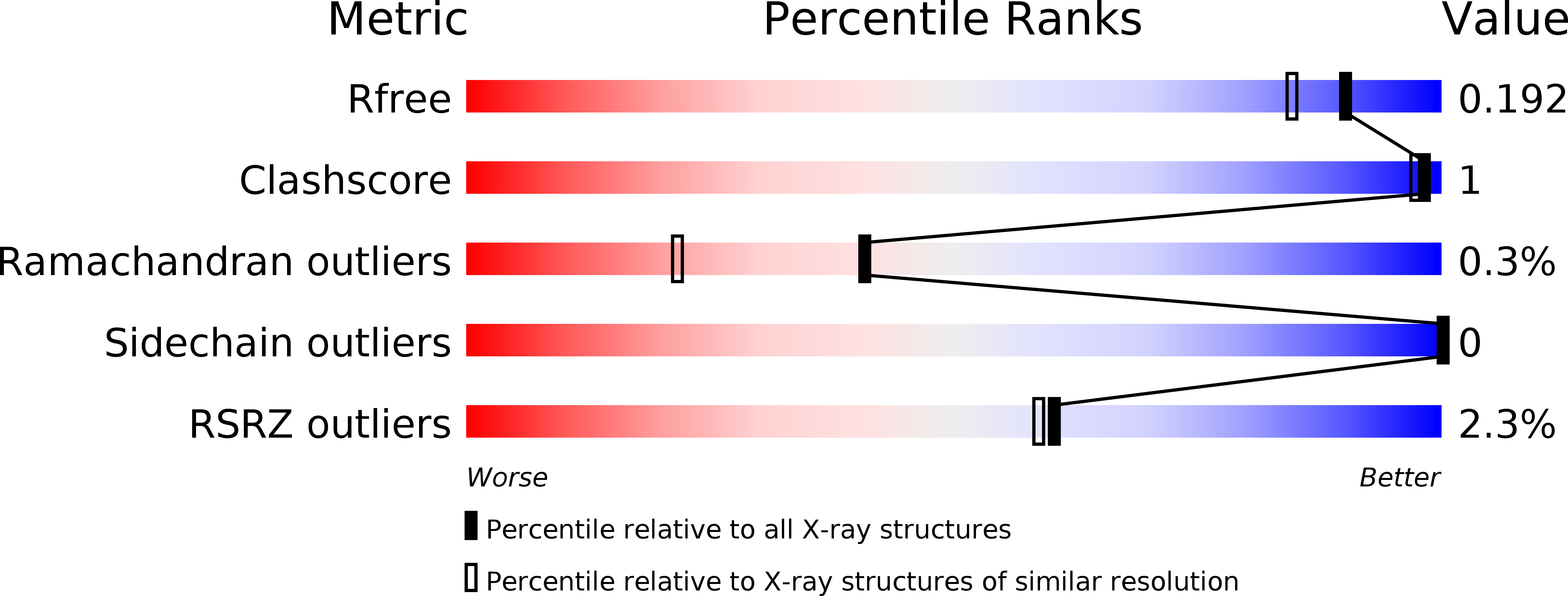
5EO8 image
Deposition Date 2015-11-10
Release Date 2016-06-29
Last Version Date 2024-03-20
Entry Detail
PDB ID:
5EO8
Title:
Crystal structure of AOL(868)
Biological Source:
Source Organism:
Host Organism:
Method Details:
Experimental Method:
Resolution:
1.60 Å
R-Value Free:
0.18
R-Value Work:
0.16
R-Value Observed:
0.16
Space Group:
P 61 2 2
Macromolecular Entities
Structures with similar UniProt ID
Protein Blast
Polymer Type:polypeptide(L)
Molecule:Predicted protein
Gene (Uniprot):fleA
Chain IDs:A
Chain Length:311
Number of Molecules:1
Biological Source:Aspergillus oryzae RIB40
Ligand Molecules
Primary Citation
Six independent fucose-binding sites in the crystal structure of Aspergillus oryzae lectin
Biochem.Biophys.Res.Commun. 477 477 482 (2016)
PMID: 27318092 DOI: 10.1016/j.bbrc.2016.06.069

Abstact

The crystal structure of AOL (a fucose-specific lectin of Aspergillus oryzae) has been solved by SAD (single-wavelength anomalous diffraction) and MAD (multi-wavelength anomalous diffraction) phasing of seleno-fucosides. The overall structure is a six-bladed β-propeller similar to that of other fucose-specific lectins. The fucose moieties of the seleno-fucosides are located in six fucose-binding sites. Although the Arg and Glu/Gln residues bound to the fucose moiety are common to all fucose-binding sites, the amino-acid residues involved in fucose binding at each site are not identical. The varying peak heights of the seleniums in the electron density map suggest that each fucose-binding site has a different carbohydrate binding affinity.

Legend

Protein

Chemical

Disease

Primary Citation of related structures