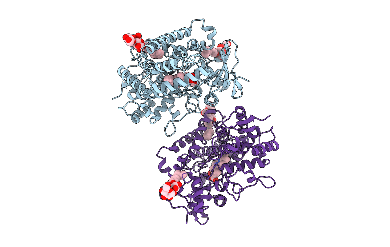
5EM4 image
Entry Detail
PDB ID:
5EM4
Keywords:
Title:
Structure of CYP2B4 F244W in a ligand free conformation
Biological Source:
Source Organism:
Host Organism:
PDB Version:
Deposition Date:
2015-11-05
Release Date:
2016-03-23
Method Details:
Experimental Method:
Resolution:
3.02 Å
R-Value Free:
0.28
R-Value Work:
0.22
R-Value Observed:
0.23
Space Group:
P 31
Macromolecular Entities
Polymer Type:polypeptide(L)
Description:Cytochrome P450 2B4
Mutations:H226Y, F244W
Chain IDs:A, B
Chain Length:492
Number of Molecules:2
Biological Source:Oryctolagus cuniculus
Primary Citation
Coumarin Derivatives as Substrate Probes of Mammalian Cytochromes P450 2B4 and 2B6: Assessing the Importance of 7-Alkoxy Chain Length, Halogen Substitution, and Non-Active Site Mutations.
Biochemistry 55 1997 2007 (2016)
PMID: 26982502 DOI: 10.1021/acs.biochem.5b01330

Abstact

Using a combined structural and biochemical approach, the functional importance of a recently described peripheral pocket bounded by the E-, F-, G-, and I-helices in CYP2B4 and 2B6 was probed. Three series of 4-substituted-7-alkoxycoumarin derivatives with -H, -CH3, or -CF3 at the 4 position of the coumarin core were used initially to monitor functional differences between CYP2B4 and 2B6. 7-Ethoxy-4-(trifluoromethyl)coumarin (7-EFC) displayed the highest catalytic efficiency among these substrates. Mutants were made to alter side-chain polarity (V/E194Q) or bulk (F/Y244W) to alter access to the peripheral pocket. Modest increases in catalytic efficiency of 7-EFC O-deethylation by the mutants were magnified considerably by chlorination or bromination of the substrate ethoxy chain. A structure of CYP2B6 Y244W in complex with (+)-α-pinene was solved at 2.2 Å and showed no CYMAL-5 in the peripheral pocket. A ligand free structure of CYP2B4 F244W was solved at 3.0 Å with CYMAL-5 in the peripheral pocket. In both instances, comparison of the respective wild-type and mutant CYP2B enzymes revealed that CYMAL-5 occupancy of the peripheral pocket had little effect on the topology of active site residue side-chains, despite the fact that the peripheral pocket and active site are located on opposite sides of the I-helix. Analysis of available CYP2B structures suggest that the effect of the amino acid substitutions within the peripheral pocket derive from altered interactions between the F and G helices.

Legend

Protein

Chemical

Disease

Primary Citation of related structures