5ELV image
Entry Detail
PDB ID:
5ELV
Title:
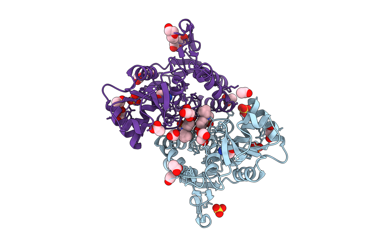
Crystal structure of the GluA2 ligand-binding domain (S1S2J-L504-N775) in complex with glutamate and BPAM-521 at 1.92 A resolution
Biological Source:
Source Organism:
PDB Version:
Deposition Date:
2015-11-05
Release Date:
2016-05-04
Method Details:
Experimental Method:
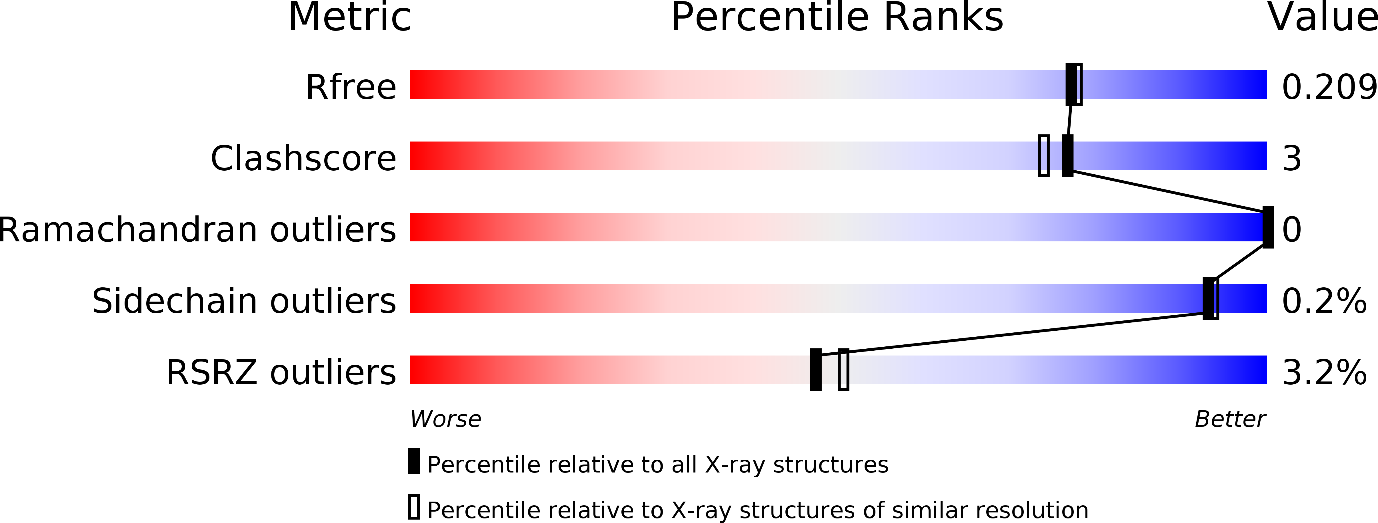
Resolution:
1.92 Å
R-Value Free:
0.20
R-Value Work:
0.16
R-Value Observed:
0.16
Space Group:
P 21 21 2
Macromolecular Entities
Polymer Type:polypeptide(L)
Description:Glutamate receptor 2,Glutamate receptor 2
Mutations:L504Y and N775S
Chain IDs:A, B
Chain Length:264
Number of Molecules:2
Biological Source:Rattus norvegicus
Primary Citation

Abstact

The 1,2,4-benzothiadiazine 1,1-dioxide type of positive allosteric modulators of the ionotropic glutamate receptor A2 (GluA2) are promising lead compounds for the treatment of cognitive disorders, e.g., Alzheimer's disease. The modulators bind in a cleft formed by the interface of two neighboring ligand binding domains and act by stabilizing the agonist-bound open-channel conformation. The driving forces behind the binding of these modulators can be significantly altered with only minor substitutions to the parent molecules. In this study, we show that changing the 7-fluorine substituent of modulators BPAM97 (2) and BPAM344 (3) into a hydroxyl group (BPAM557 (4) and BPAM521 (5), respectively), leads to a more favorable binding enthalpy (ΔH, kcal/mol) from -4.9 (2) and -7.5 (3) to -6.2 (4) and -14.5 (5), but also a less favorable binding entropy (-TΔS, kcal/mol) from -2.3 (2) and -1.3 (3) to -0.5 (4) and 4.8 (5). Thus, the dissociation constants (Kd, μM) of 4 (11.2) and 5 (0.16) are similar to those of 2 (5.6) and 3 (0.35). Functionally, 4 and 5 potentiated responses of 10 μM L-glutamate at homomeric rat GluA2(Q)i receptors with EC50 values of 67.3 and 2.45 μM, respectively. The binding mode of 5 was examined with x-ray crystallography, showing that the only change compared to that of earlier compounds was the orientation of Ser-497 pointing toward the hydroxyl group of 5. The favorable enthalpy can be explained by the formation of a hydrogen bond from the side-chain hydroxyl group of Ser-497 to the hydroxyl group of 5, whereas the unfavorable entropy might be due to desolvation effects combined with a conformational restriction of Ser-497 and 5. In summary, this study shows a remarkable example of enthalpy-entropy compensation in drug development accompanied with a likely explanation of the underlying structural mechanism.

Legend

Protein

Chemical

Disease

Primary Citation of related structures