5EKG image
Deposition Date 2015-11-03
Release Date 2016-02-10
Last Version Date 2024-03-06
Entry Detail
PDB ID:
5EKG
Keywords:
Title:
Crystallization and X-ray Diffraction Data Collection of Importin-alpha from Mus musculus Complexed with a XPG NLS Peptide, fragment 2
Biological Source:
Source Organism:
Host Organism:
Method Details:
Experimental Method:
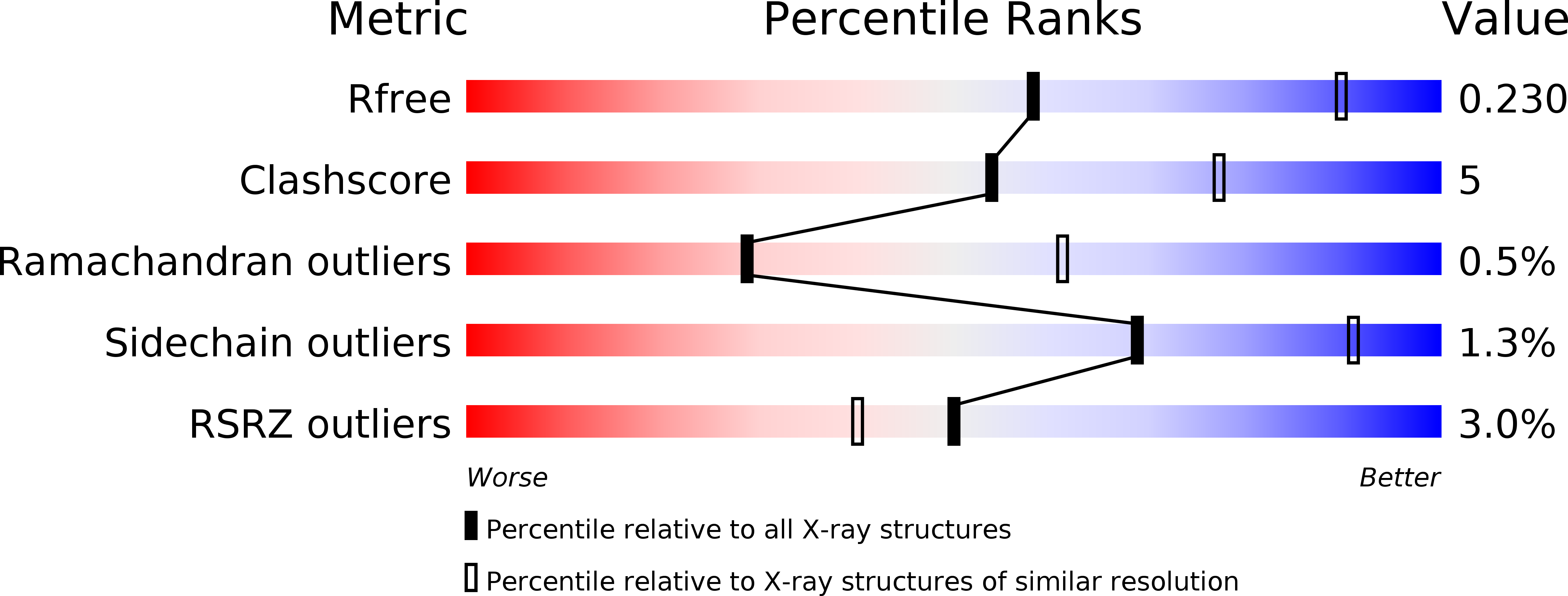
Resolution:
2.80 Å
R-Value Free:
0.22
R-Value Work:
0.18
R-Value Observed:
0.18
Space Group:
P 21 21 21
Macromolecular Entities
Polymer Type:polypeptide(L)
Molecule:Importin subunit alpha-1
Gene (Uniprot):Kpna2
Chain IDs:C (auth: A)
Chain Length:510
Number of Molecules:1
Biological Source:Mus musculus
Polymer Type:polypeptide(L)
Molecule:XPG2 peptide
Chain IDs:A (auth: B), B (auth: C)
Chain Length:19
Number of Molecules:2
Biological Source:synthetic construct
Ligand Molecules
Primary Citation
Structural and Calorimetric Studies Demonstrate that Xeroderma Pigmentosum Type G (XPG) Can Be Imported to the Nucleus by a Classical Nuclear Import Pathway via a Monopartite NLS Sequence.
J.Mol.Biol. 428 2120 2131 (2016)
PMID: 26812207 DOI: 10.1016/j.jmb.2016.01.019

Abstact

Xeroderma pigmentosum type G (XPG) proteins are involved in DNA lesion recognition and promotion of nucleotide excision repair. Specific mutations in these proteins may lead to Cockayne syndrome, in which the patients may display severe developmental retardation and neurological abnormalities. No structural information is available for their spacer region or the C-terminal domain, which are important, respectively, for specific nucleotide excision repair activity and substrate specificity, as well as nuclear translocation. Immunofluorescence studies suggested two specific regions of the XPG C-terminus as potential bipartite nuclear localization sequences, which would be responsible for its translocation to the nucleus by the classical nuclear import pathway mediated by the importin-α (Impα). Thus, in order to test these hypotheses and gain insight into the structural basis for the nuclear import process for the XPG protein, we solved the crystal structures of complexes formed by the Impα and peptides corresponding to both putative nuclear localization signal (NLS) sequences (XPG1 and XPG2) and performed isothermal titration calorimetry assays to determine their binding affinities. Structural experiments confirm the binding of both NLS peptides to Impα but, unexpectedly, they bind to the receptor as monopartite NLSs. The isothermal titration calorimetry assays demonstrated that XPG1 and XPG2 peptides bind to two separate binding sites, but with high affinity to the major NLS-binding site of the Impα, resembling classical monopartite SV40 TAg NLS. The results lead to insights about what distinguishes monopartite and bipartite NLSs, as well as the differential roles of XPG1 and XPG2 NLSs in the nuclear localization of XPG.

Legend

Protein

Chemical

Disease

Primary Citation of related structures