5EHA image
Entry Detail
PDB ID:
5EHA
Keywords:
Title:
Crystal structure of recombinant MtaL at 1.35 Angstrom resolution
Biological Source:
Source Organism:
Host Organism:
PDB Version:
Deposition Date:
2015-10-28
Release Date:
2016-03-09
Method Details:
Experimental Method:
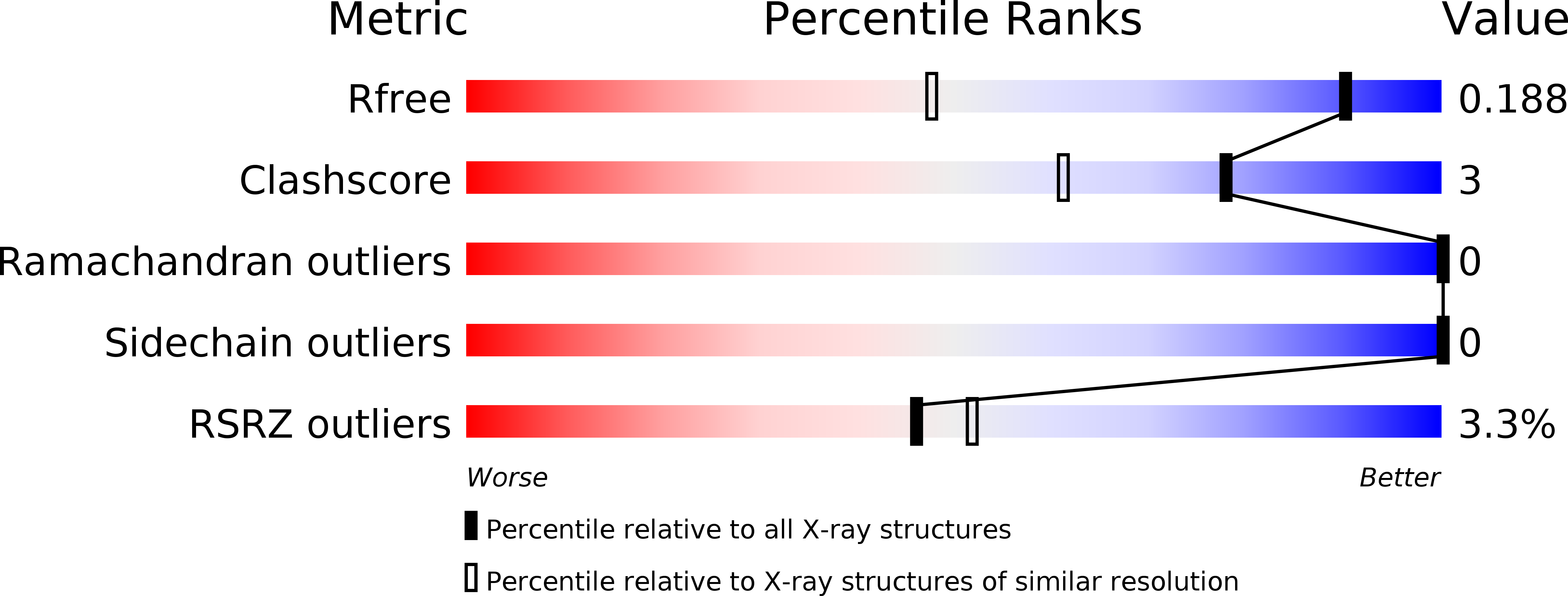
Resolution:
1.35 Å
R-Value Free:
0.18
R-Value Work:
0.16
R-Value Observed:
0.16
Space Group:
I 1 2 1
Macromolecular Entities
Polymer Type:polypeptide(L)
Description:Lectin-like fold protein
Chain IDs:A
Chain Length:153
Number of Molecules:1
Biological Source:Agaricus bisporus
Primary Citation
Crystal structure of recombinant tyrosinase-binding protein MtaL at 1.35 angstrom resolution.
Acta Crystallogr.,Sect.F 72 244 250 (2016)
PMID: 26919530 DOI: 10.1107/S2053230X16002107

Abstact

Mushroom tyrosinase-associated lectin-like protein (MtaL) binds to mature Agaricus bisporus tyrosinase in vivo, but the exact physiological function of MtaL is unknown. In this study, the crystal structure of recombinant MtaL is reported at 1.35 Å resolution. Comparison of its structure with that of the truncated and cleaved MtaL present in the complex with tyrosinase directly isolated from mushroom shows that the general β-trefoil fold is conserved. However, differences are detected in the loop regions, particularly in the β2-β3 loop, which is intact and not cleaved in the recombinant MtaL. Furthermore, the N-terminal tail is rotated inwards, covering the tyrosinase-binding interface. Thus, MtaL must undergo conformational changes in order to bind mature mushroom tyrosinase. Very interestingly, the β-trefoil fold has been identified to be essential for carbohydrate interaction in other lectin-like proteins. Comparison of the structures of MtaL and a ricin-B-like lectin with a bound disaccharide shows that MtaL may have a similar carbohydrate-binding site that might be involved in glycoreceptor activity.

Legend

Protein

Chemical

Disease

Primary Citation of related structures