Deposition Date
2015-10-28
Release Date
2015-12-23
Last Version Date
2023-09-27
Entry Detail
PDB ID:
5EH6
Keywords:
Title:
Crystal Structure of the Glycophorin A Transmembrane Monomer in Lipidic Cubic Phase
Biological Source:
Source Organism(s):
Homo sapiens (Taxon ID: 9606)
Expression System(s):
Method Details:
Experimental Method:
Resolution:
1.92 Å
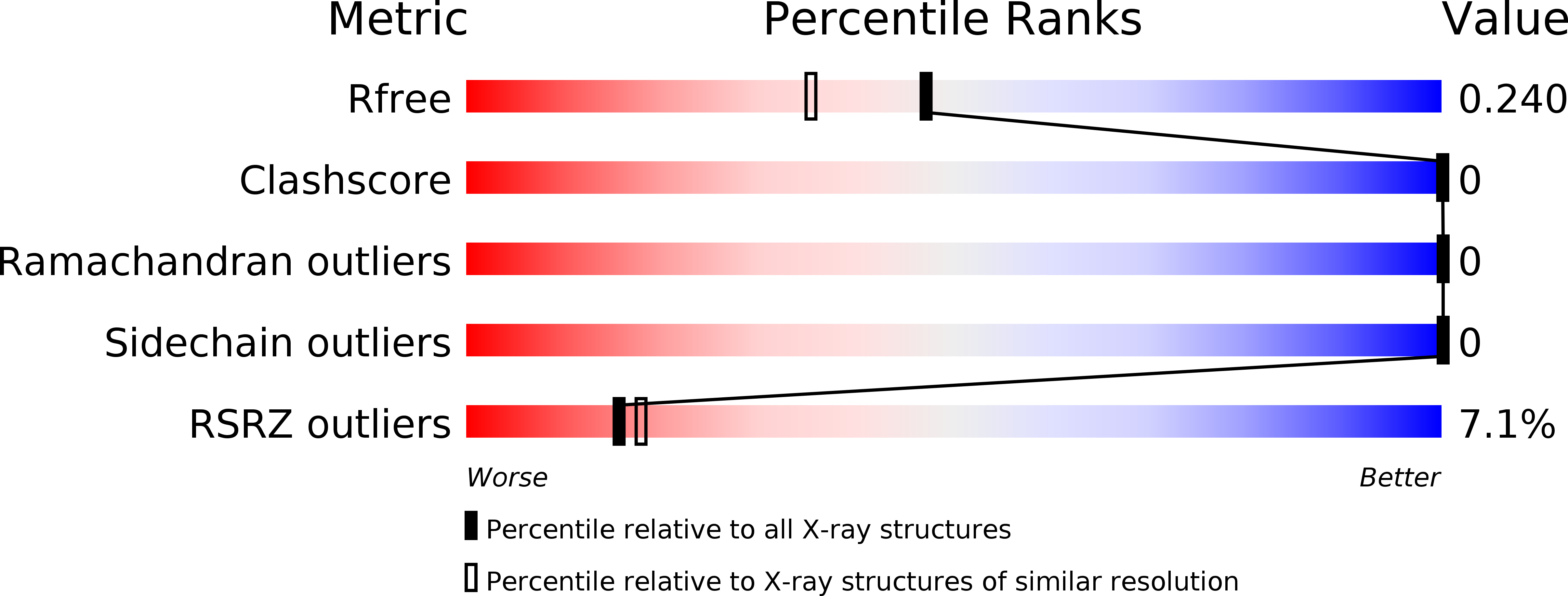
R-Value Free:
0.23
R-Value Work:
0.22
R-Value Observed:
0.22
Space Group:
H 3 2