Deposition Date
2015-10-26
Release Date
2016-05-25
Last Version Date
2024-05-08
Entry Detail
PDB ID:
5EFV
Keywords:
Title:
The host-recognition device of Staphylococcus aureus phage Phi11
Biological Source:
Source Organism(s):
Staphylococcus phage phi11 (Taxon ID: 12360)
Expression System(s):
Method Details:
Experimental Method:
Resolution:
2.20 Å
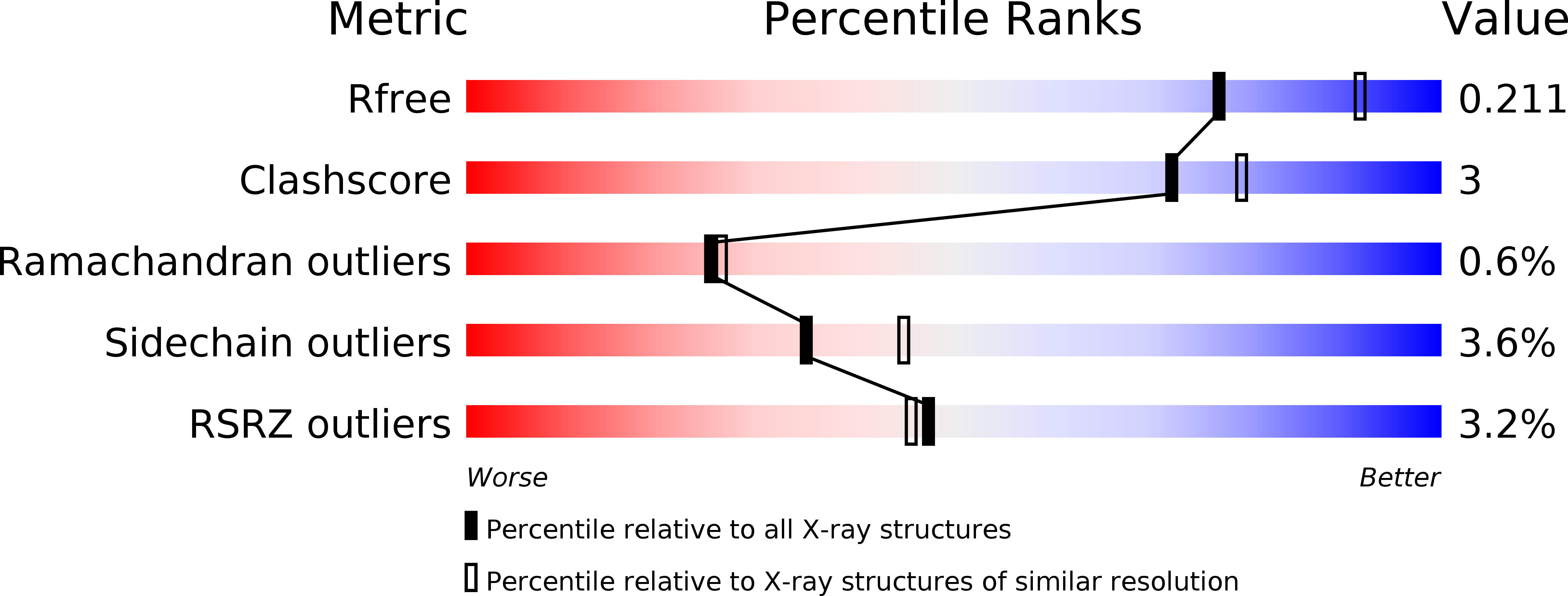
R-Value Free:
0.21
R-Value Work:
0.17
R-Value Observed:
0.17
Space Group:
P 1