
Deposition Date
2015-10-22
Release Date
2016-01-20
Last Version Date
2024-11-20
Entry Detail
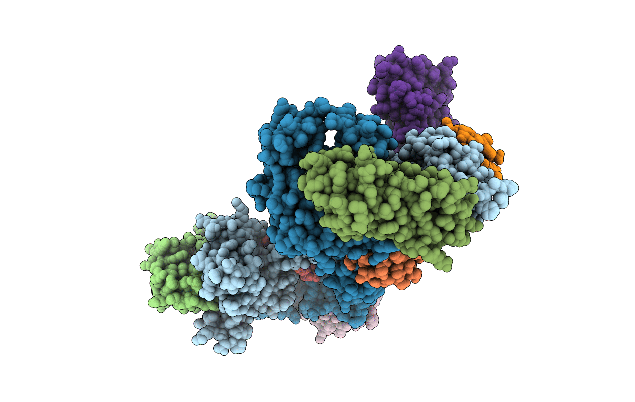
PDB ID:
5EDV
Keywords:
Title:
Structure of the HOIP-RBR/UbcH5B~ubiquitin transfer complex
Biological Source:
Source Organism(s):
Homo sapiens (Taxon ID: 9606)
Expression System(s):
Method Details:
Experimental Method:
Resolution:
3.48 Å
R-Value Free:
0.30
R-Value Work:
0.24
R-Value Observed:
0.25
Space Group:
P 1 21 1
