5ECW image
Deposition Date 2015-10-20
Release Date 2016-02-17
Last Version Date 2024-05-08
Entry Detail
PDB ID:
5ECW
Keywords:
Title:
Structure of the Shigella flexneri VapC mutant D7A
Biological Source:
Source Organism:
Method Details:
Experimental Method:
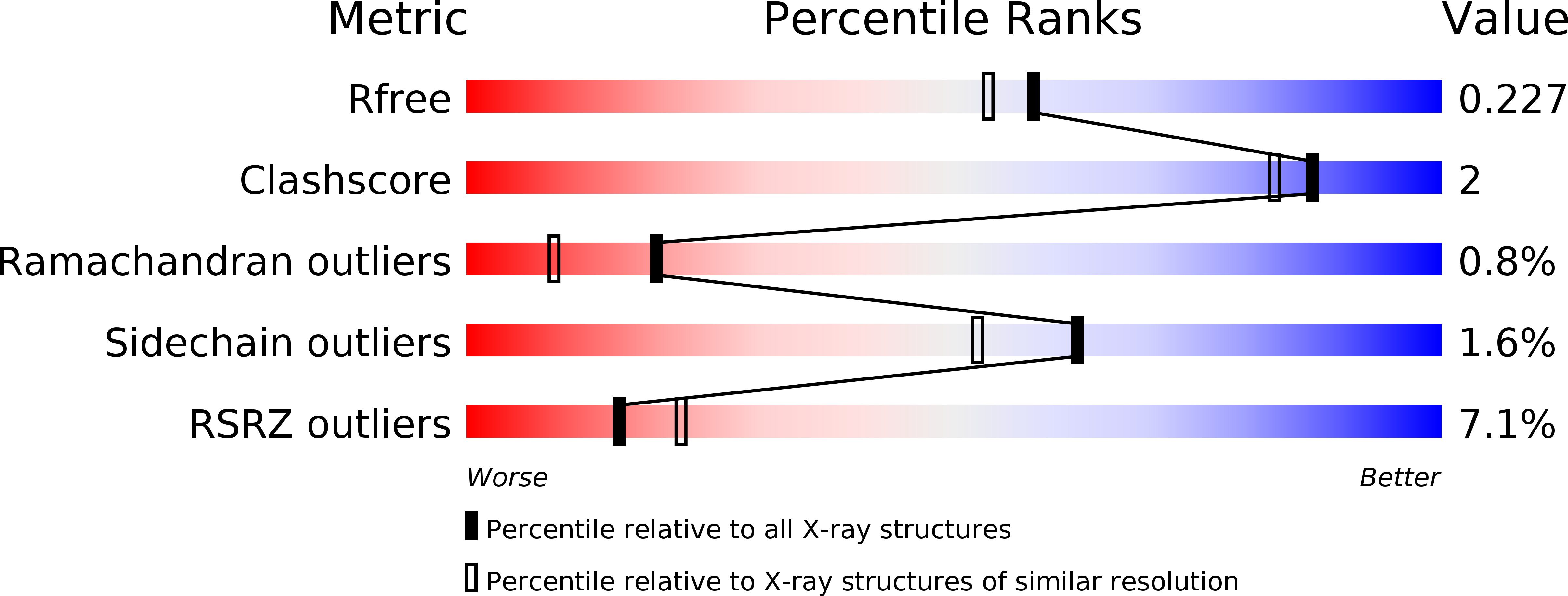
Resolution:
1.94 Å
R-Value Free:
0.22
R-Value Work:
0.18
R-Value Observed:
0.19
Space Group:
H 3
Macromolecular Entities
Polymer Type:polypeptide(L)
Molecule:tRNA(fMet)-specific endonuclease VapC
Gene (Uniprot):vapC
Mutations:D7A
Chain IDs:A, B
Chain Length:138
Number of Molecules:2
Biological Source:Shigella flexneri
Primary Citation
Structural analysis of the active site architecture of the VapC toxin from Shigella flexneri.
Proteins 84 892 899 (2016)
PMID: 26833558 DOI: 10.1002/prot.25002

Abstact

The VapC toxin from the Shigella flexneri 2a virulence plasmid pMYSH6000 belongs to the PIN domain protein family, which is characterized by a conserved fold with low amino acid sequence conservation. The toxin is a bona fide Mg(2+) -dependent ribonuclease and has been shown to target initiator tRNA(fMet) in vivo. Here, we present crystal structures of active site catalytic triad mutants D7A, D7N, and D98N of the VapC toxin in absence of antitoxin. In all structures, as well as in solution, VapC forms a dimer. In the D98N structure, a Hepes molecule occupies both active sites of the dimer and comparison with the structure of RNase H bound to a DNA/RNA hybrid suggests that the Hepes molecule mimics the position of an RNA nucleotide in the VapC active site. Proteins 2016; 84:892-899. © 2016 Wiley Periodicals, Inc.

Legend

Protein

Chemical

Disease

Primary Citation of related structures