5EBX image
Deposition Date 1989-12-20
Release Date 1990-04-15
Last Version Date 2024-11-20
Entry Detail
PDB ID:
5EBX
Keywords:
Title:
THE CRYSTAL STRUCTURE OF ERABUTOXIN A AT 2.0 ANGSTROMS RESOLUTION
Biological Source:
Source Organism:
Method Details:
Experimental Method:
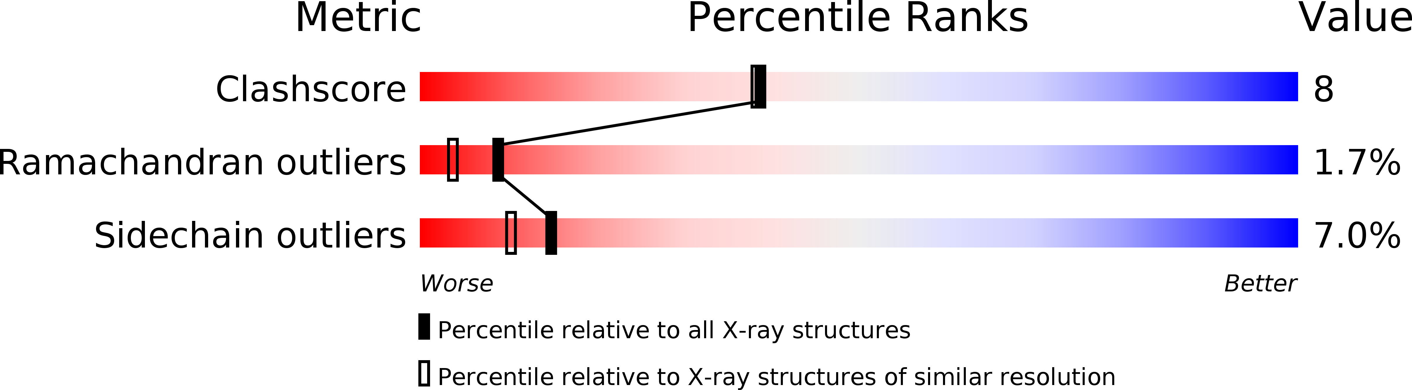
Resolution:
2.00 Å
R-Value Observed:
0.16
Space Group:
P 21 21 21
Macromolecular Entities
Structures with similar UniProt ID
Protein Blast
Polymer Type:polypeptide(L)
Molecule:ERABUTOXIN A
Chain IDs:A
Chain Length:62
Number of Molecules:1
Biological Source:Laticauda semifasciata
Ligand Molecules
Primary Citation

Abstact

The three-dimensional structure of erabutoxin a, a single-chain, 62-residue protein neurotoxin from snake venom, has been determined to 2.0-A resolution by x-ray crystal structure analysis. Molecular replacement methods were used, and the structure refined to a residual R = 0.17. The sites of 62 water molecules and 1 sulfate ion have been located and refined. The structure of erabutoxin a is very similar to that established earlier for erabutoxin b. These toxins from venom of the same snake differ in sequence only at residue 26, which is Asn in erabutoxin a and His in erabutoxin b. The substitution leads to only minor variations in intramolecular hydrogen bonding. Furthermore, the distribution of thermal parameters and the implied regional mobilities are similar in the two structures. In particular, the highly mobile character of the peripheral segment Pro44-Gly49 in both structures supports the specific role proposed for this segment in neurotoxin binding to the acetylcholine receptor. Forty-eight of the solvent sites determined are first surface positions; approximately one-half of these are equivalent to solvent sites in erabutoxin b.

Legend

Protein

Chemical

Disease

Primary Citation of related structures
Feedback Form
Name
Email
Institute
Feedback