Deposition Date
2015-10-19
Release Date
2016-02-17
Last Version Date
2024-11-20
Entry Detail
PDB ID:
5EBC
Keywords:
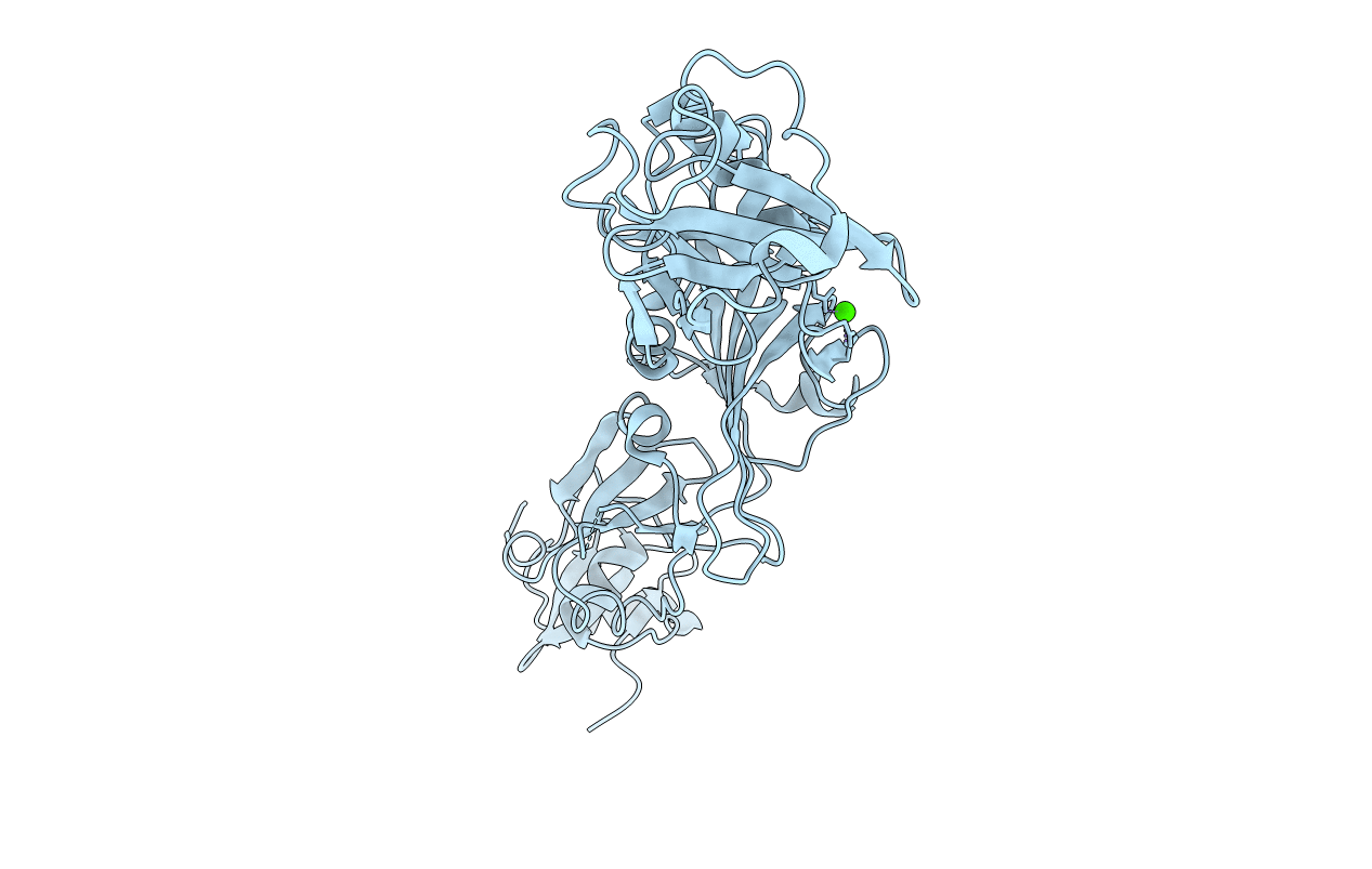
Title:
Crystal structure of EccB1 of Mycobacterium tuberculosis in spacegroup P21 (state III)
Biological Source:
Source Organism(s):
Mycobacterium tuberculosis (Taxon ID: 83332)
Expression System(s):
Method Details:
Experimental Method:
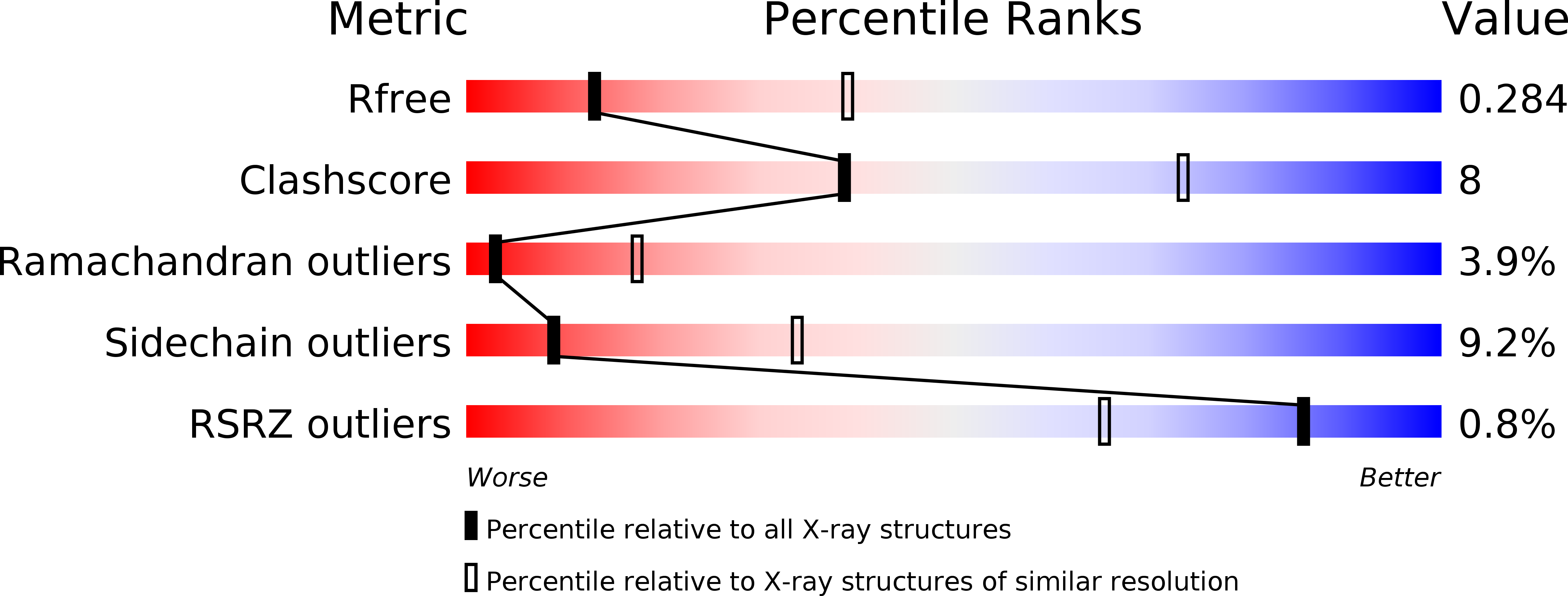
Resolution:
3.00 Å
R-Value Free:
0.28
R-Value Work:
0.23
R-Value Observed:
0.23
Space Group:
P 1 21 1