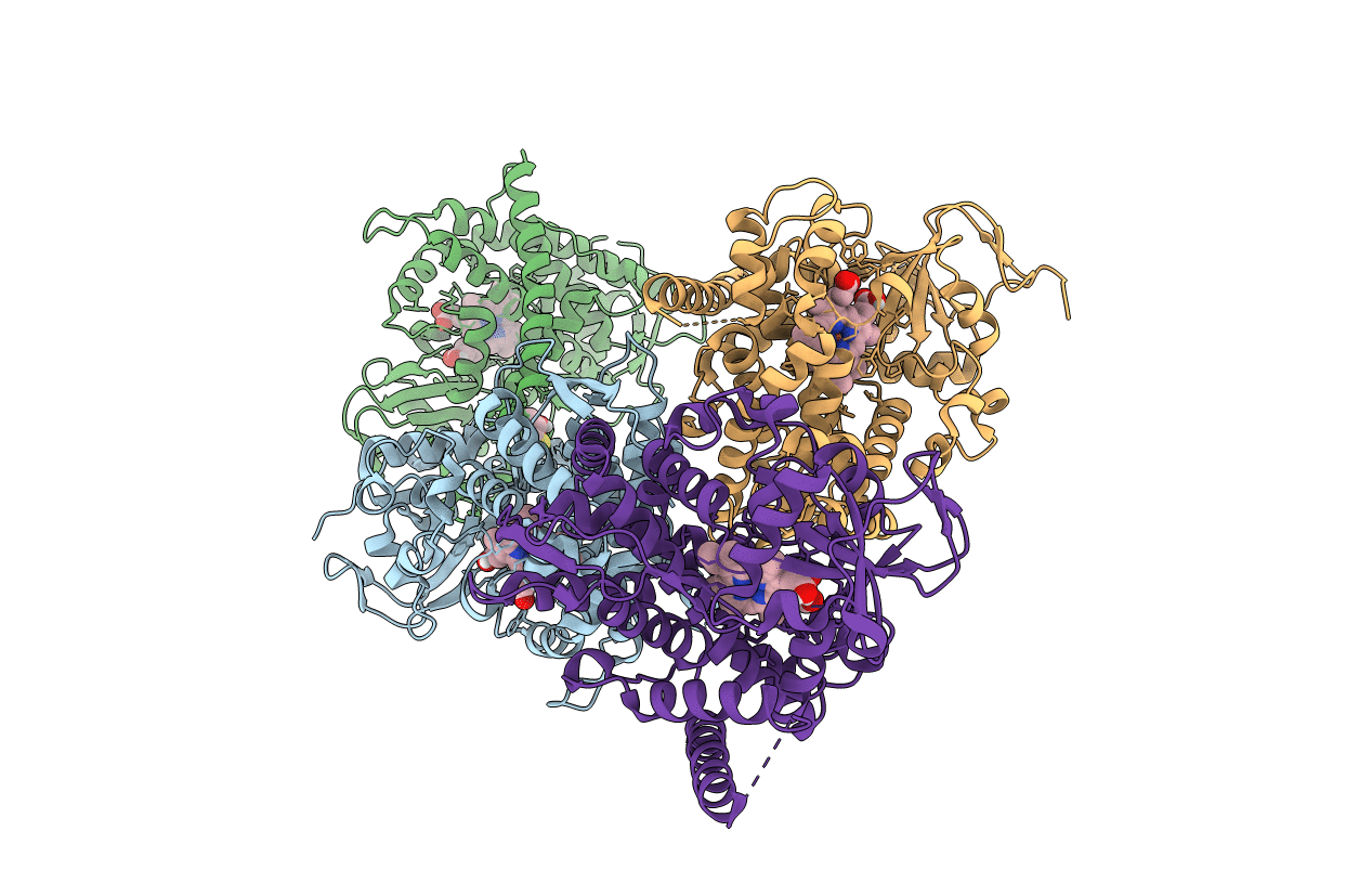
5E9Z image
Entry Detail
PDB ID:
5E9Z
Keywords:
Title:
Cytochrome P450 BM3 mutant M11
Biological Source:
Source Organism:
Host Organism:
PDB Version:
Deposition Date:
2015-10-15
Release Date:
2016-01-27
Method Details:
Experimental Method:
Resolution:
2.23 Å
R-Value Free:
0.22
R-Value Work:
0.18
R-Value Observed:
0.18
Space Group:
C 1 2 1
Macromolecular Entities
Polymer Type:polypeptide(L)
Description:Bifunctional cytochrome P450/NADPH--P450 reductase
Mutations:R47L, E64G, F81I, F87V, E143G, L188Q, Y198C, E267V, H285Y, G415S
Chain IDs:A, B, C, D
Chain Length:469
Number of Molecules:4
Biological Source:Bacillus megaterium
Primary Citation
Insights into regioselective metabolism of mefenamic acid by cytochrome P450 BM3 mutants through crystallography, docking, molecular dynamics, and free energy calculations.
Proteins 84 383 396 (2016)
PMID: 26757175 DOI: 10.1002/prot.24985

Abstact

Cytochrome P450 BM3 (CYP102A1) mutant M11 is able to metabolize a wide range of drugs and drug-like compounds. Among these, M11 was recently found to be able to catalyze formation of human metabolites of mefenamic acid and other nonsteroidal anti-inflammatory drugs (NSAIDs). Interestingly, single active-site mutations such as V87I were reported to invert regioselectivity in NSAID hydroxylation. In this work, we combine crystallography and molecular simulation to study the effect of single mutations on binding and regioselective metabolism of mefenamic acid by M11 mutants. The heme domain of the protein mutant M11 was expressed, purified, and crystallized, and its X-ray structure was used as template for modeling. A multistep approach was used that combines molecular docking, molecular dynamics (MD) simulation, and binding free-energy calculations to address protein flexibility. In this way, preferred binding modes that are consistent with oxidation at the experimentally observed sites of metabolism (SOMs) were identified. Whereas docking could not be used to retrospectively predict experimental trends in regioselectivity, we were able to rank binding modes in line with the preferred SOMs of mefenamic acid by M11 and its mutants by including protein flexibility and dynamics in free-energy computation. In addition, we could obtain structural insights into the change in regioselectivity of mefenamic acid hydroxylation due to single active-site mutations. Our findings confirm that use of MD and binding free-energy calculation is useful for studying biocatalysis in those cases in which enzyme binding is a critical event in determining the selective metabolism of a substrate.

Legend

Protein

Chemical

Disease

Primary Citation of related structures