Deposition Date
2015-10-15
Release Date
2016-06-08
Last Version Date
2023-09-27
Entry Detail
PDB ID:
5E9E
Keywords:
Title:
Crystal Structure of the Alpha-kinase Domain of Myosin-II Heavy Chain Kinase A in Complex with AMP-PNP
Biological Source:
Source Organism(s):
Dictyostelium discoideum (Taxon ID: 44689)
Expression System(s):
Method Details:
Experimental Method:
Resolution:
2.40 Å
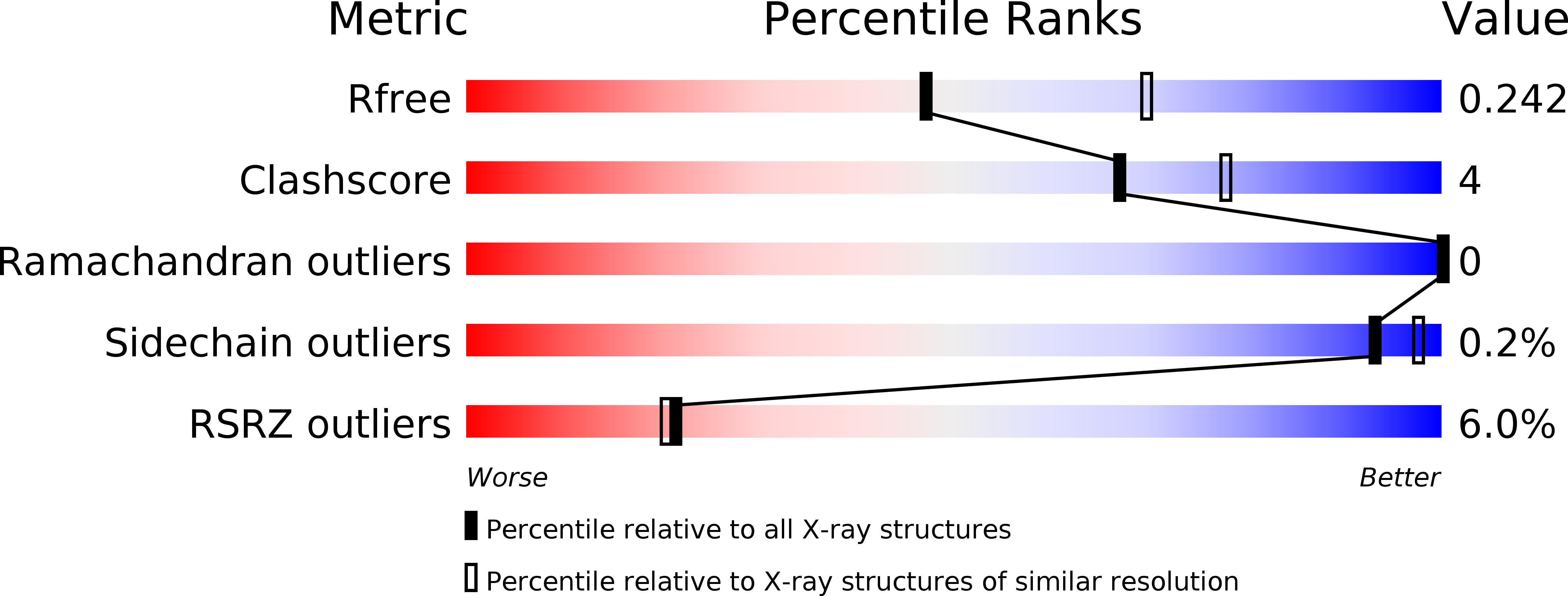
R-Value Free:
0.23
R-Value Work:
0.19
R-Value Observed:
0.19
Space Group:
P 21 21 2