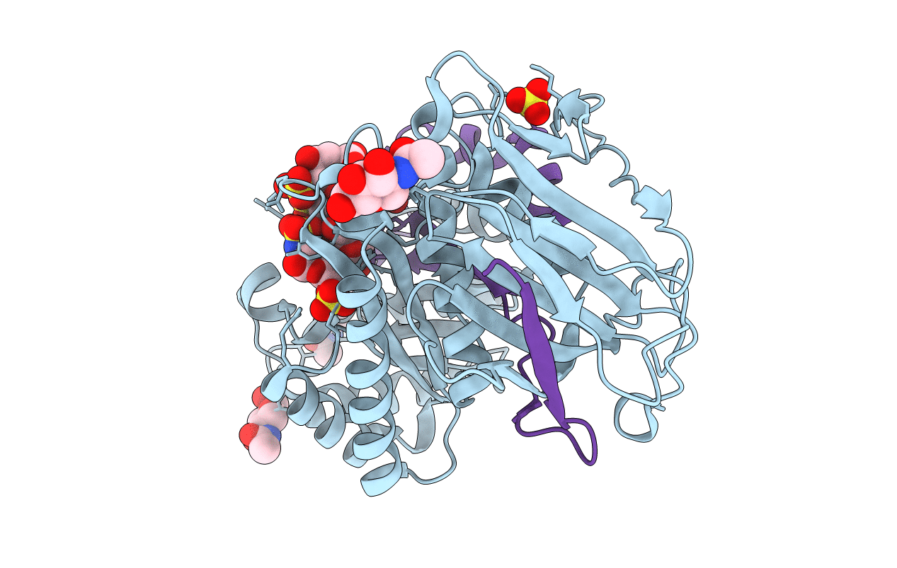
5E9C image
Deposition Date 2015-10-15
Release Date 2015-11-18
Last Version Date 2024-11-06
Entry Detail
PDB ID:
5E9C
Keywords:
Title:
Crystal structure of human heparanase in complex with heparin tetrasaccharide dp4
Biological Source:
Source Organism:
Homo sapiens (Taxon ID: 9606)
Host Organism:
Method Details:
Experimental Method:
Resolution:
1.73 Å
R-Value Free:
0.21
R-Value Work:
0.17
R-Value Observed:
0.17
Space Group:
P 1 21 1
Macromolecular Entities
Polymer Type:polypeptide(L)
Molecule:Heparanase
Gene (Uniprot):HPSE
Chain IDs:A
Chain Length:389
Number of Molecules:1
Biological Source:Homo sapiens
Polymer Type:polypeptide(L)
Molecule:Heparanase
Gene (Uniprot):HPSE
Chain IDs:B
Chain Length:77
Number of Molecules:1
Biological Source:Homo sapiens
Primary Citation
Structural characterization of human heparanase reveals insights into substrate recognition.
Nat.Struct.Mol.Biol. 22 1016 1022 (2015)
PMID: 26575439 DOI: 10.1038/nsmb.3136

Abstact

Heparan sulfate (HS) is a glycosaminoglycan that forms a key component of the extracellular matrix (ECM). Breakdown of HS is carried out by heparanase (HPSE), an endo-β-glucuronidase of the glycoside hydrolase 79 (GH79) family. Overexpression of HPSE results in breakdown of extracellular HS and release of stored growth factors and hence is strongly linked to cancer metastasis. Here we present crystal structures of human HPSE at 1.6-Å to 1.9-Å resolution that reveal how an endo-acting binding cleft is exposed by proteolytic activation of latent proHPSE. We used oligosaccharide complexes to map the substrate-binding and sulfate-recognition motifs. These data shed light on the structure and interactions of a key enzyme involved in ECM maintenance and provide a starting point for the design of HPSE inhibitors for use as biochemical tools and anticancer therapeutics.

Legend

Protein

Chemical

Disease

Primary Citation of related structures