5E96 image
Deposition Date 2015-10-14
Release Date 2016-07-06
Last Version Date 2023-09-27
Entry Detail
PDB ID:
5E96
Keywords:
Title:
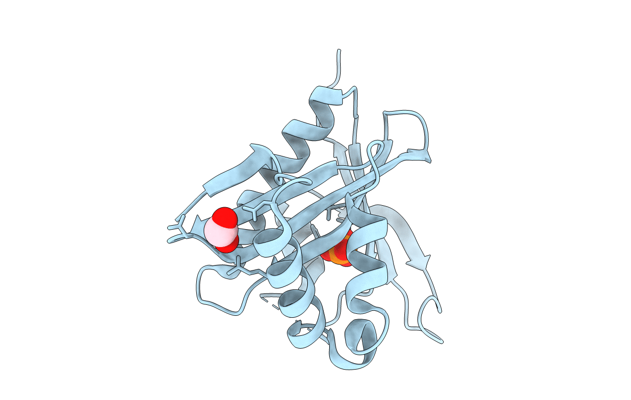
Crystal structure of aminoglycoside 6'-acetyltransferase type Ii
Biological Source:
Source Organism:
Host Organism:
Method Details:
Experimental Method:
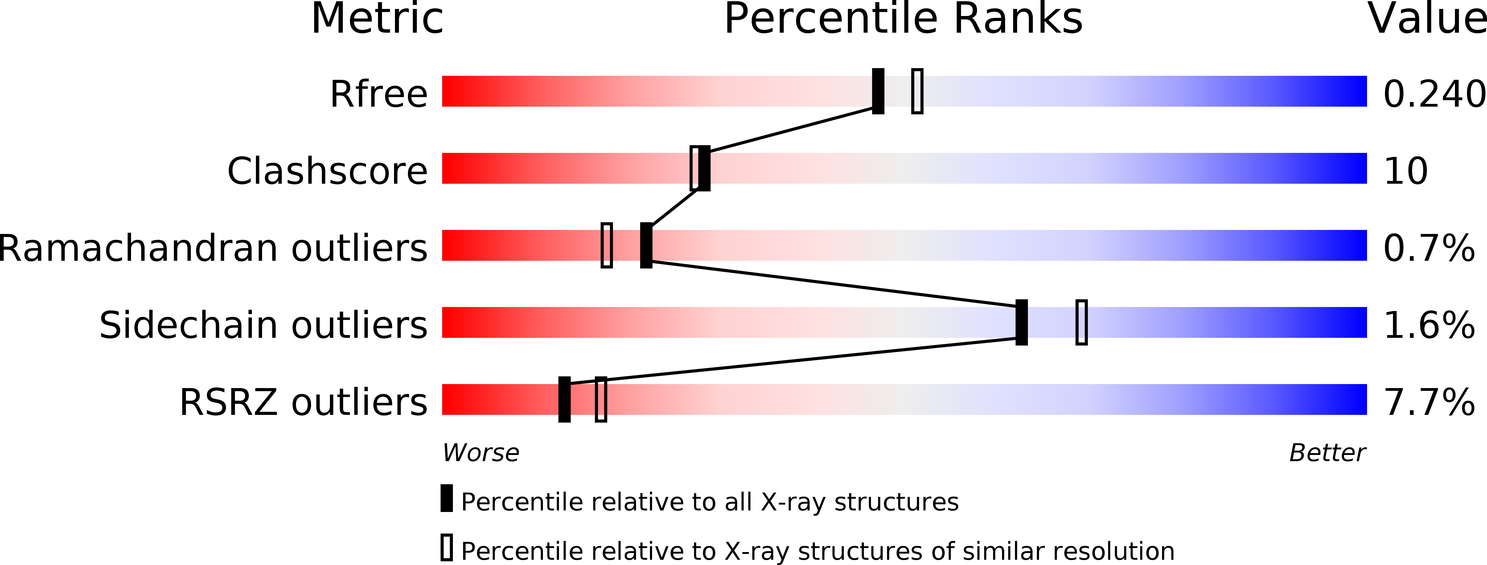
Resolution:
2.10 Å
R-Value Free:
0.25
R-Value Work:
0.21
R-Value Observed:
0.21
Space Group:
P 64
Macromolecular Entities
Structures with similar UniProt ID
Protein Blast
Polymer Type:polypeptide(L)
Molecule:Aminoglycoside 6'-acetyltransferase
Chain IDs:A
Chain Length:180
Number of Molecules:1
Biological Source:Enterococcus faecium
Primary Citation
Comprehensive characterization of ligand-induced plasticity changes in a dimeric enzyme.
Febs J. 283 3029 3038 (2016)
PMID: 27333541 DOI: 10.1111/febs.13788

Abstact

UNLABELLED An enzyme's inherent structural plasticity is frequently associated with substrate binding, yet detailed structural characterization of flexible proteins remains challenging. This study employs complementary biophysical methods to characterize the partially unfolded structure of substrate-free AAC(6')-Ii, an N-acetyltransferase of the GCN5-related N-acetyltransferase (GNAT) superfamily implicated in conferring broad-spectrum aminoglycoside resistance on Enterococcus faecium. The X-ray crystal structure of AAC(6')-Ii is analyzed to identify relative motions of the structural elements that constitute the dimeric enzyme. Comparison with the previously elucidated crystal structure of AAC(6')-Ii with acetyl coenzyme A (AcCoA) reveals conformational changes that occur upon substrate binding. Our understanding of the enzyme's structural plasticity is further refined with small-angle X-ray scattering and circular dichroism analyses, which together reveal how flexible structural elements impact dimerization and substrate binding. These results clarify the extent of unfolding that AAC(6')-Ii undergoes in the absence of AcCoA and provide a structural connection to previously observed allosteric cooperativity of this enzyme. DATABASE Structural data are available in the PDB database under the accession number 5E96.

Legend

Protein

Chemical

Disease

Primary Citation of related structures
Feedback Form
Name
Email
Institute
Feedback