5E8I image
Deposition Date 2015-10-14
Release Date 2015-12-09
Last Version Date 2023-09-27
Entry Detail
PDB ID:
5E8I
Title:
Crystal structure of the DNA binding domain of human transcription factor FLI1 in complex with a 10-mer DNA ACCGGAAGTG
Biological Source:
Source Organism:
Homo sapiens (Taxon ID: 9606)
Endothia gyrosa (Taxon ID: 40263)
Host Organism:
Method Details:
Experimental Method:
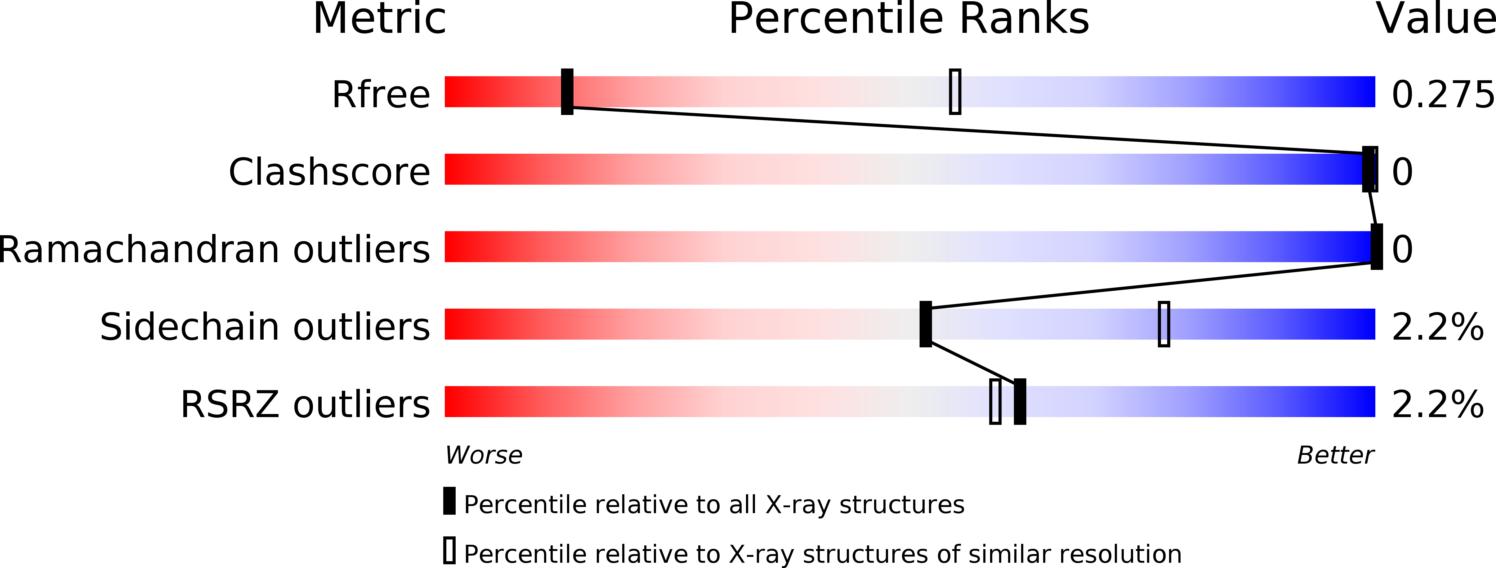
Resolution:
3.45 Å
R-Value Free:
0.27
R-Value Work:
0.22
R-Value Observed:
0.22
Space Group:
P 31 2 1
Macromolecular Entities
Polymer Type:polypeptide(L)
Molecule:Friend leukemia integration 1 transcription factor
Gene (Uniprot):FLI1
Chain IDs:A, D, G, J
Chain Length:128
Number of Molecules:4
Biological Source:Homo sapiens
Polymer Type:polydeoxyribonucleotide
Molecule:DNA (5'-D(*AP*CP*CP*GP*GP*AP*AP*GP*TP*G)-3')
Chain IDs:B, E, H, K
Chain Length:10
Number of Molecules:4
Biological Source:Endothia gyrosa
Polymer Type:polydeoxyribonucleotide
Molecule:DNA (5'-D(*CP*AP*CP*TP*TP*CP*CP*GP*GP*T)-3')
Chain IDs:C, F, I, L
Chain Length:10
Number of Molecules:4
Biological Source:Endothia gyrosa
Ligand Molecules
Primary Citation
Structural Basis for Dimerization and DNA Binding of Transcription Factor FLI1.
Biochemistry 54 7365 7374 (2015)
PMID: 26618620 DOI: 10.1021/acs.biochem.5b01121

Abstact

FLI1 (Friend leukemia integration 1) is a metazoan transcription factor that is upregulated in a number of cancers. In addition, rearrangements of the fli1 gene cause sarcomas, leukemias, and lymphomas. These rearrangements encode oncogenic transcription factors, in which the DNA binding domain (DBD or ETS domain) of FLI1 on the C-terminal side is fused to a part of an another protein on the N-terminal side. Such abnormal cancer cell-specific fusions retain the DNA binding properties of FLI1 and acquire non-native protein-protein or protein-nucleic acid interactions of the substituted region. As a result, these fusions trigger oncogenic transcriptional reprogramming of the host cell. Interactions of FLI1 fusions with other proteins and with itself play a critical role in the oncogenic regulatory functions, and they are currently under intense scrutiny, mechanistically and as potential novel anticancer drug targets. We report elusive crystal structures of the FLI1 DBD, alone and in complex with cognate DNA containing a GGAA recognition sequence. Both structures reveal a previously unrecognized dimer of this domain, consistent with its dimerization in solution. The homodimerization interface is helix-swapped and dominated by hydrophobic interactions, including those between two interlocking Phe362 residues. A mutation of Phe362 to an alanine disrupted the propensity of this domain to dimerize without perturbing its structure or the DNA binding function, consistent with the structural observations. We propose that FLI1 DBD dimerization plays a role in transcriptional activation and repression by FLI1 and its fusions at promoters containing multiple FLI1 binding sites.

Legend

Protein

Chemical

Disease

Primary Citation of related structures