5E8F image
Deposition Date 2015-10-14
Release Date 2015-11-18
Last Version Date 2024-01-10
Entry Detail
PDB ID:
5E8F
Keywords:
Title:
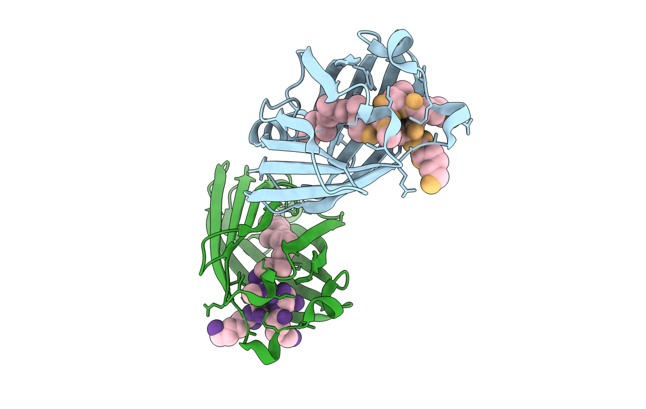
Structure of Fully modified geranylgeranylated PDE6C Peptide in complex with PDE6D
Biological Source:
Source Organism:
Homo sapiens (Taxon ID: 9606)
Host Organism:
Method Details:
Experimental Method:
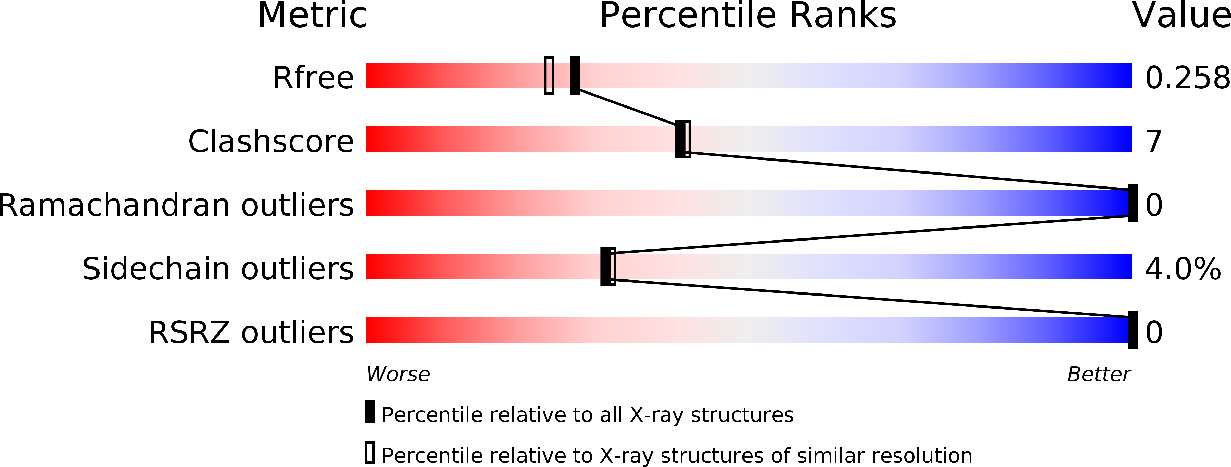
Resolution:
2.10 Å
R-Value Free:
0.25
R-Value Work:
0.19
R-Value Observed:
0.20
Space Group:
C 2 2 21
Macromolecular Entities
Polymer Type:polypeptide(L)
Molecule:Retinal rod rhodopsin-sensitive cGMP 3',5'-cyclic phosphodiesterase subunit delta
Gene (Uniprot):PDE6D
Chain IDs:A, B (auth: C)
Chain Length:149
Number of Molecules:2
Biological Source:Homo sapiens
Polymer Type:polypeptide(L)
Molecule:Cone cGMP-specific 3',5'-cyclic phosphodiesterase subunit alpha'
Gene (Uniprot):PDE6C
Chain IDs:C (auth: D), D (auth: E)
Chain Length:5
Number of Molecules:2
Biological Source:Homo sapiens
Modified Residue
Compound ID Chain ID Parent Comp ID Details 2D Image
CMT C CYS modified residue
Ligand Molecules
Primary Citation
The N- and C-terminal ends of RPGR can bind to PDE6 delta.
Embo Rep. 16 1583 1585 (2015)
PMID: 26553937 DOI: 10.15252/embr.201541404

Abstact

This study shows that the prenylated C‐terminus of RPGR can bind to PDE6δ with high affinity, suggesting two distinct binding sites of the RPGR/PDE6δ complex. The serine residue at the −3 position relative to the prenylated cysteine seems to play a key role in defining the selectivity of PDE6δ towards ciliary prenylated cargo. [Image: see text]

Legend

Protein

Chemical

Disease

Primary Citation of related structures