5E8E image
Entry Detail
PDB ID:
5E8E
Title:
Crystal structure of thrombin bound to an exosite 1-specific IgA Fab
Biological Source:
Source Organism:
PDB Version:
Deposition Date:
2015-10-14
Release Date:
2015-10-28
Method Details:
Experimental Method:
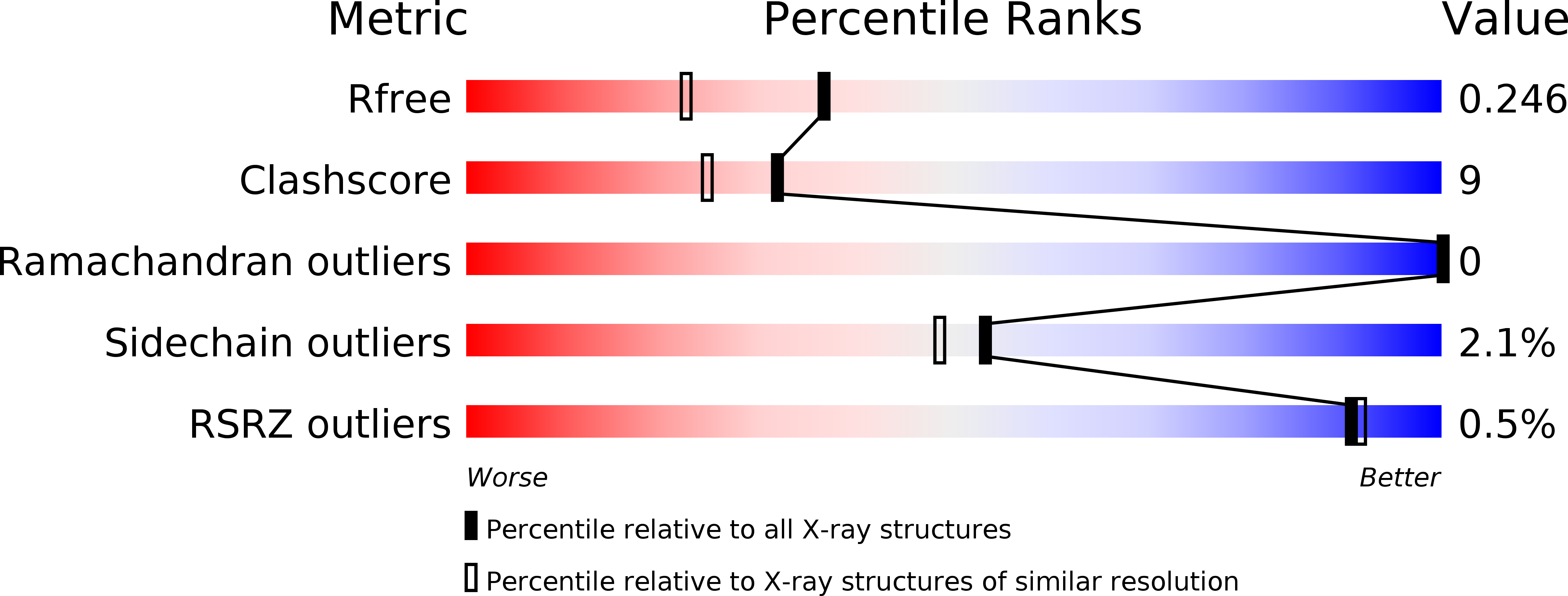
Resolution:
1.90 Å
R-Value Free:
0.24
R-Value Work:
0.20
R-Value Observed:
0.20
Space Group:
P 21 21 21
Macromolecular Entities
Polymer Type:polypeptide(L)
Description:IGA FAB LIGHT CHAIN
Chain IDs:A
Chain Length:215
Number of Molecules:1
Biological Source:Homo sapiens
Polymer Type:polypeptide(L)
Description:IGA FAB HEAVY CHAIN
Chain IDs:B
Chain Length:229
Number of Molecules:1
Biological Source:Homo sapiens
Polymer Type:polypeptide(L)
Description:THROMBIN HEAVY CHAIN
Chain IDs:C (auth: H)
Chain Length:259
Number of Molecules:1
Biological Source:Homo sapiens
Polymer Type:polypeptide(L)
Description:THROMBIN LIGHT CHAIN
Chain IDs:D (auth: L)
Chain Length:36
Number of Molecules:1
Biological Source:Homo sapiens
Modified Residue
Compound ID Chain ID Parent Comp ID Details 2D Image
ASN A ASN GLYCOSYLATION SITE
PCA B GLU PYROGLUTAMIC ACID
Peptide-like Molecules
PRD_000020
Primary Citation
Discovery and characterization of an antibody directed against exosite I of thrombin.
J.Thromb.Haemost. 14 137 142 (2016)
PMID: 26469093 DOI: 10.1111/jth.13171

Abstact

UNLABELLED ESSENTIALS: An IgA paraprotein with anti-thrombin activity was not associated with a severe bleeding phenotype. This observation challenges the paradigm that anticoagulant therapy necessarily increases bleeding risk. Characterization of the antibody showed that it specifically binds to thrombin exosite I. A therapeutic drug with the properties of this antibody might be an antithrombotic that doesn't cause bleeding. BACKGROUND We report the case of a 54-year-old female who presented with a traumatic subdural hemorrhage. Coagulation tests were markedly prolonged due to the presence of an anti-thrombin IgA paraprotein at 3 g L(-1) . The patient made a complete recovery and has had no abnormal bleeding during a 7-year follow-up, despite the persistence of the paraprotein. OBJECTIVES To determine how the paraprotein prolonged clotting tests by defining its target and its epitope. METHODS The paraprotein was purified and added to normal pooled plasma for in vitro clotting assays. Binding studies were conducted to determine the affinity of the IgA for thrombin. The Fab was isolated and crystallized with thrombin. RESULTS The purified IgA was sufficient to confer the patient's in vitro coagulation profile in normal pooled plasma, and was found to bind specifically and with high affinity to thrombin. A crystal structure of the Fab fragment in complex with thrombin revealed an exosite I interaction involving CDRH3 of the antibody. CONCLUSIONS Although the patient originally presented with a subdural bleed, the hematoma resolved without intervention, and no other bleeding event occurred during the subsequent 7 years. During this period, the patient's IgA paraprotein levels have remained constant at 3 g L(-1) , suggesting that the presence of a high-affinity, exosite I-directed antibody is consistent with normal hemostasis. A therapeutic derivative of this antibody might therefore permit antithrombotic dose escalation without an associated increase in the risk of bleeding.

Legend

Protein

Chemical

Disease

Primary Citation of related structures