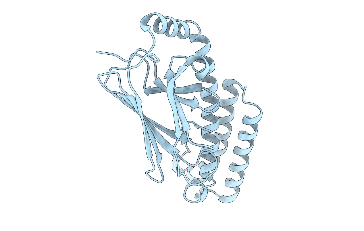
5E86 image
Deposition Date 2015-10-13
Release Date 2015-12-30
Last Version Date 2024-03-06
Entry Detail
PDB ID:
5E86
Keywords:
Title:
isolated SBD of BiP with loop34 modification
Biological Source:
Source Organism:
Homo sapiens (Taxon ID: 9606)
Host Organism:
Method Details:
Experimental Method:
Resolution:
2.68 Å
R-Value Free:
0.25
R-Value Work:
0.21
R-Value Observed:
0.21
Space Group:
C 2 2 21
Macromolecular Entities
Polymer Type:polypeptide(L)
Molecule:78 kDa glucose-regulated protein
Gene (Uniprot):HSPA5
Mutations:loop34 modification
Chain IDs:A
Chain Length:232
Number of Molecules:1
Biological Source:Homo sapiens
Primary Citation
Close and Allosteric Opening of the Polypeptide-Binding Site in a Human Hsp70 Chaperone BiP.
Structure 23 2191 2203 (2015)
PMID: 26655470 DOI: 10.1016/j.str.2015.10.012

Abstact

Binding immunoglobulin protein (BiP), an essential and ubiquitous Hsp70 chaperone in the ER, plays a key role in protein folding and quality control. BiP contains two functional domains: a nucleotide-binding domain (NBD) and a substrate-binding domain (SBD). NBD binds and hydrolyzes ATP; the substrates for SBD are extended polypeptides. ATP binding allosterically accelerates polypeptide binding and release. Although crucial to the chaperone activity, the molecular mechanisms of polypeptide binding and allosteric coupling of BiP are poorly understood. Here, we present crystal structures of an intact human BiP in the ATP-bound state, the first intact eukaryotic Hsp70 structure, and isolated BiP-SBD with a peptide substrate bound representing the ADP-bound state. These structures and our biochemical analysis demonstrate that BiP has a unique NBD-SBD interface that is highly conserved only in eukaryotic Hsp70s found in the cytosol and ER to fortify its ATP-bound state and promote the opening of its polypeptide-binding pocket.

Legend

Protein

Chemical

Disease

Primary Citation of related structures