Deposition Date
2015-10-12
Release Date
2016-09-28
Last Version Date
2024-01-10
Entry Detail
PDB ID:
5E78
Keywords:
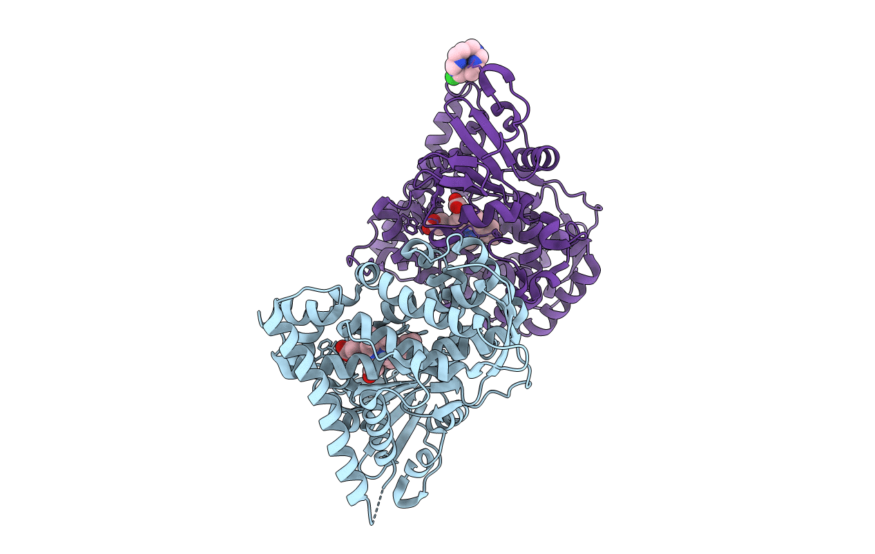
Title:
Crystal structure of P450 BM3 heme domain variant complexed with Co(III)Sep
Biological Source:
Source Organism(s):
Bacillus megaterium (Taxon ID: 1404)
Expression System(s):
Method Details:
Experimental Method:
Resolution:
2.00 Å
R-Value Free:
0.21
R-Value Work:
0.17
R-Value Observed:
0.17
Space Group:
P 21 21 21