5E70 image
Deposition Date 2015-10-11
Release Date 2015-12-16
Last Version Date 2023-09-27
Entry Detail
PDB ID:
5E70
Keywords:
Title:
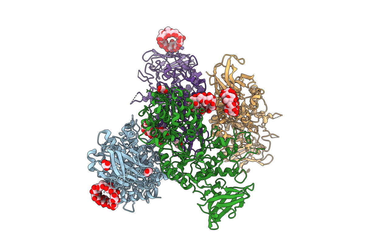
Crystal structure of Ecoli Branching Enzyme with gamma cyclodextrin
Biological Source:
Host Organism:
Method Details:
Experimental Method:
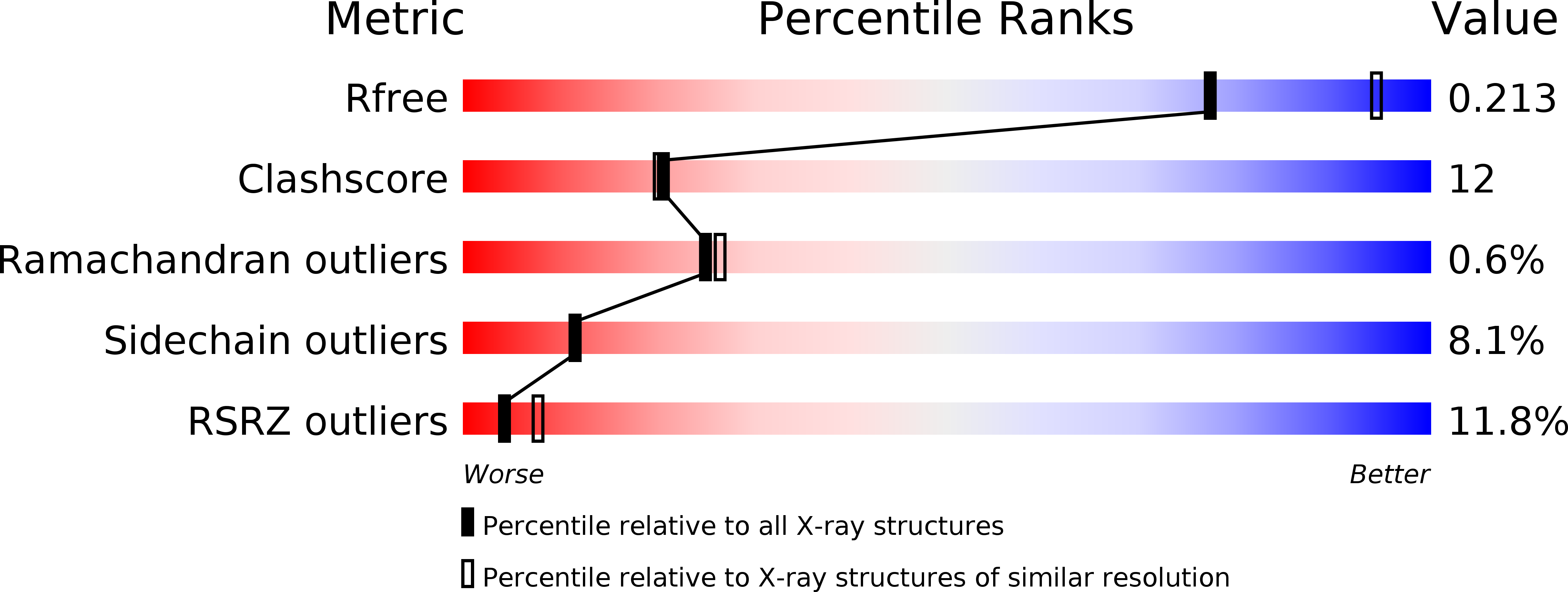
Resolution:
2.33 Å
R-Value Free:
0.21
R-Value Work:
0.17
R-Value Observed:
0.17
Space Group:
P 1 21 1
Macromolecular Entities
Polymer Type:polypeptide(L)
Molecule:1,4-alpha-glucan branching enzyme GlgB
Gene (Uniprot):glgB
Chain IDs:A, B, C, D
Chain Length:612
Number of Molecules:4
Biological Source:Escherichia coli O139:H28 (strain E24377A / ETEC)
Ligand Molecules
Peptide-like Molecules
PRD_900042
Primary Citation
Crystal structures of Escherichia coli branching enzyme in complex with cyclodextrins.
Acta Crystallogr D Struct Biol 72 641 647 (2016)
PMID: 27139627 DOI: 10.1107/S2059798316003272

Abstact

Branching enzyme (BE) is responsible for the third step in glycogen/starch biosynthesis. It catalyzes the cleavage of α-1,4 glucan linkages and subsequent reattachment to form α-1,6 branch points. These branches are crucial to the final structure of glycogen and starch. The crystal structures of Escherichia coli BE (EcBE) in complex with α-, β- and γ-cyclodextrin were determined in order to better understand substrate binding. Four cyclodextrin-binding sites were identified in EcBE; they were all located on the surface of the enzyme, with none in the vicinity of the active site. While three of the sites were also identified as linear polysaccharide-binding sites, one of the sites is specific for cyclodextrins. In previous work three additional binding sites were identified as exclusively binding linear malto-oligosaccharides. Comparison of the binding sites shed light on this apparent specificity. Binding site IV is located in the carbohydrate-binding module 48 (CBM48) domain of EcBE and superimposes with the cyclodextrin-binding site found in the CBM48 domain of 5'-AMP-activated protein kinase (AMPK). Comparison of these sites shows the similarities and differences in the two binding modes. While some of the binding sites were found to be conserved between branching enzymes of different organisms, some are quite divergent, indicating both similarities and differences between oligosaccharide binding in branching enzymes from various sources.

Legend

Protein

Chemical

Disease

Primary Citation of related structures