5E6N image
Deposition Date 2015-10-10
Release Date 2016-01-06
Last Version Date 2024-03-20
Entry Detail
PDB ID:
5E6N
Keywords:
Title:
Crystal structure of C. elegans LGG-2
Biological Source:
Source Organism:
Host Organism:
Method Details:
Experimental Method:
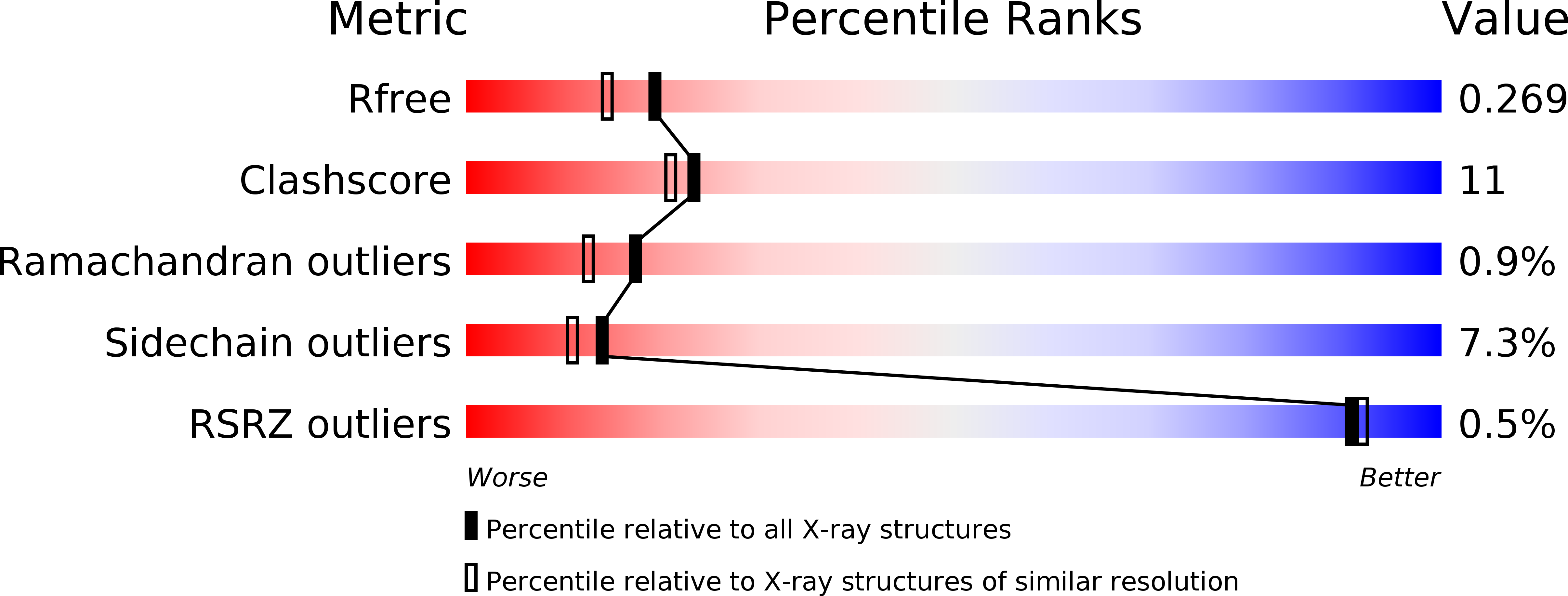
Resolution:
2.10 Å
R-Value Free:
0.26
R-Value Work:
0.21
R-Value Observed:
0.22
Space Group:
C 1 2 1
Macromolecular Entities
Polymer Type:polypeptide(L)
Molecule:Protein lgg-2
Gene (Uniprot):lgg-2
Chain IDs:A, B
Chain Length:118
Number of Molecules:2
Biological Source:Caenorhabditis elegans
Primary Citation
Structural Basis of the Differential Function of the Two C. elegans Atg8 Homologs, LGG-1 and LGG-2, in Autophagy
Mol.Cell 60 914 929 (2015)
PMID: 26687600 DOI: 10.1016/j.molcel.2015.11.019

Abstact

Multicellular organisms have multiple homologs of the yeast ATG8 gene, but the differential roles of these homologs in autophagy during development remain largely unknown. Here we investigated structure/function relationships in the two C. elegans Atg8 homologs, LGG-1 and LGG-2. lgg-1 is essential for degradation of protein aggregates, while lgg-2 has cargo-specific and developmental-stage-specific roles in aggregate degradation. Crystallography revealed that the N-terminal tails of LGG-1 and LGG-2 adopt the closed and open form, respectively. LGG-1 and LGG-2 interact differentially with autophagy substrates and Atg proteins, many of which carry a LIR motif. LGG-1 and LGG-2 have structurally distinct substrate binding pockets that prefer different residues in the interacting LIR motif, thus influencing binding specificity. Lipidated LGG-1 and LGG-2 possess distinct membrane tethering and fusion activities, which may result from the N-terminal differences. Our study reveals the differential function of two ATG8 homologs in autophagy during C. elegans development.

Legend

Protein

Chemical

Disease

Primary Citation of related structures