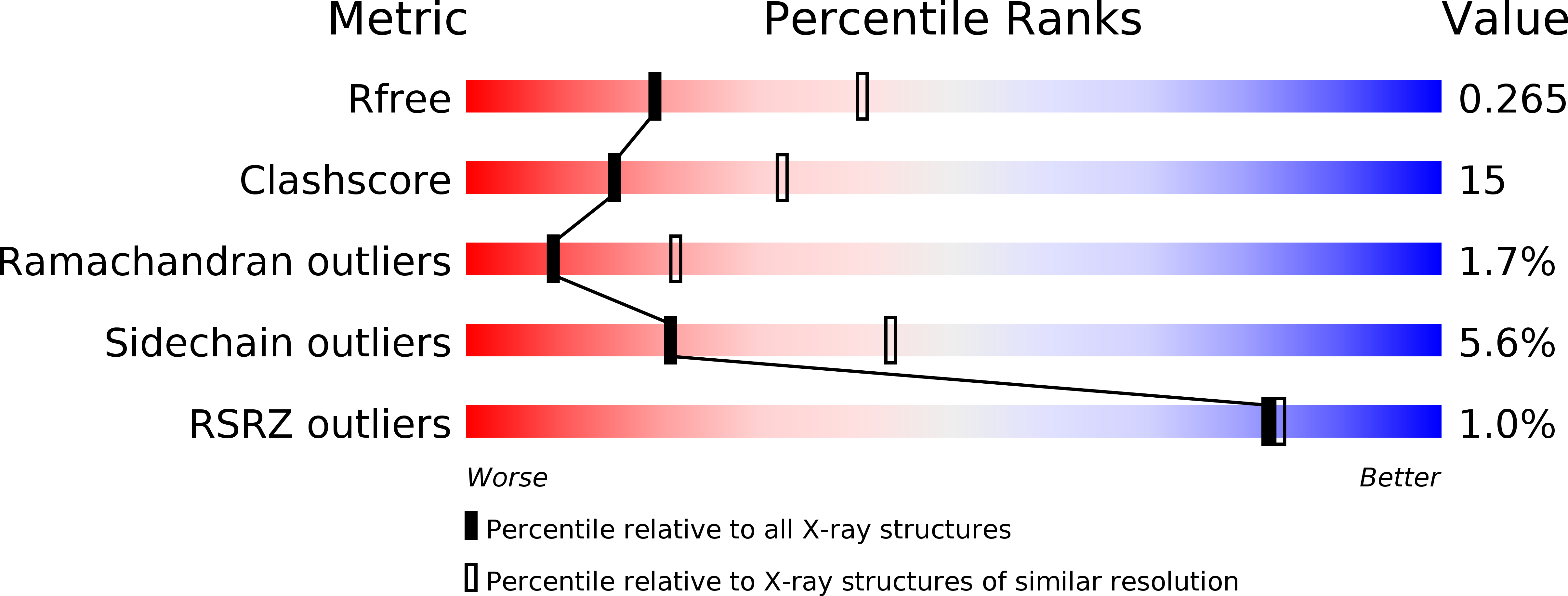
5E4V image
Deposition Date 2015-10-07
Release Date 2016-01-27
Last Version Date 2024-01-10
Entry Detail
PDB ID:
5E4V
Keywords:
Title:
Crystal structure of measles N0-P complex
Biological Source:
Method Details:
Experimental Method:
Resolution:
2.71 Å
R-Value Free:
0.26
R-Value Work:
0.21
R-Value Observed:
0.21
Space Group:
P 31 2 1
Macromolecular Entities
Structures with similar UniProt ID
Protein Blast
Polymer Type:polypeptide(L)
Molecule:Nucleoprotein,Phosphoprotein
Gene (Uniprot):P
Chain IDs:A
Chain Length:437
Number of Molecules:1
Biological Source:Measles virus (strain Edmonston B), Measles virus (strain Edmonston-Moraten vaccine)
Primary Citation
Crystal Structure of the Measles Virus Nucleoprotein Core in Complex with an N-Terminal Region of Phosphoprotein.
J.Virol. 90 2849 2857 (2015)
PMID: 26719278 DOI: 10.1128/JVI.02865-15

Abstact

UNLABELLED The enveloped negative-stranded RNA virus measles virus (MeV) is an important human pathogen. The nucleoprotein (N(0)) assembles with the viral RNA into helical ribonucleocapsids (NC) which are, in turn, coated by a helical layer of the matrix protein. The viral polymerase complex uses the NC as its template. The N(0) assembly onto the NC and the activity of the polymerase are regulated by the viral phosphoprotein (P). In this study, we pulled down an N(0)₁₋₄₀₈ fragment lacking most of its C-terminal tail domain by several affinity-tagged, N-terminal P fragments to map the N(0)-binding region of P to the first 48 amino acids. We showed biochemically and using P mutants the importance of the hydrophobic interactions for the binding. We fused an N(0) binding peptide, P₁₋₄₈, to the C terminus of an N(0)₂₁₋₄₀₈ fragment lacking both the N-terminal peptide and the C-terminal tail of N protein to reconstitute and crystallize the N(0)-P complex. We solved the X-ray structure of the resulting N(0)-P chimeric protein at a resolution of 2.7 Å. The structure reveals the molecular details of the conserved N(0)-P interface and explains how P chaperones N(0), preventing both self-assembly of N(0) and its binding to RNA. Finally, we propose a model for a preinitiation complex for RNA polymerization. IMPORTANCE Measles virus is an important, highly contagious human pathogen. The nucleoprotein N binds only to viral genomic RNA and forms the helical ribonucleocapsid that serves as a template for viral replication. We address how N is regulated by another protein, the phosphoprotein (P), to prevent newly synthesized N from binding to cellular RNA. We describe the atomic model of an N-P complex and compare it to helical ribonucleocapsid. We thus provide insight into how P chaperones N and helps to start viral RNA synthesis. Our results provide a new insight into mechanisms of paramyxovirus replication. New data on the mechanisms of phosphoprotein chaperone action allows better understanding of virus genome replication and nucleocapsid assembly. We describe a conserved structural interface for the N-P interaction which could be a target for drug development to treat not only measles but also potentially other paramyxovirus diseases.

Legend

Protein

Chemical

Disease

Primary Citation of related structures