5E3L image
Deposition Date 2015-10-03
Release Date 2016-03-23
Last Version Date 2024-03-06
Entry Detail
PDB ID:
5E3L
Title:
Crystal structure of Fis bound to 27bp DNA F1-8G (AAATTGGTTTGAATTTTGAGCCAATTT)
Biological Source:
Source Organism:
Host Organism:
Method Details:
Experimental Method:
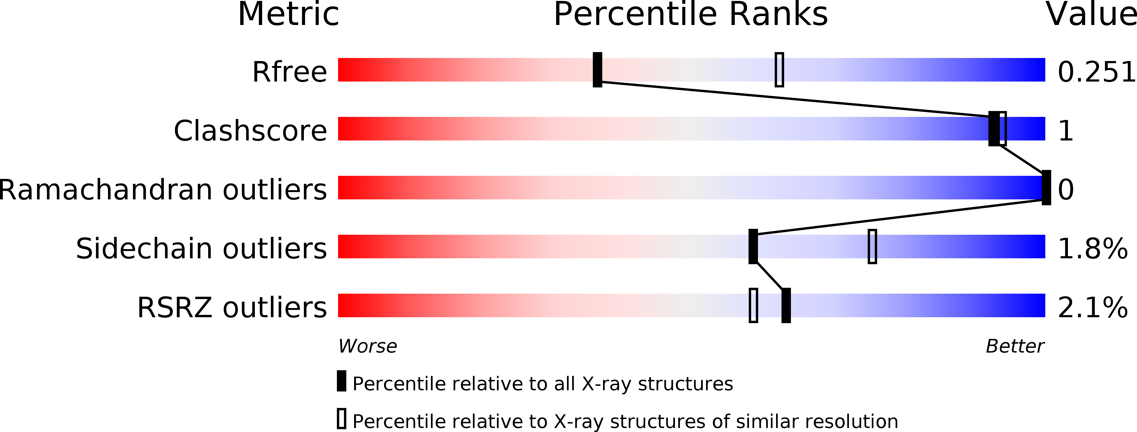
Resolution:
2.66 Å
R-Value Free:
0.24
R-Value Work:
0.20
R-Value Observed:
0.21
Space Group:
P 21 21 21
Macromolecular Entities
Structures with similar UniProt ID
Protein Blast
Polymer Type:polypeptide(L)
Molecule:DNA-binding protein Fis
Gene (Uniprot):fis
Chain IDs:A, B
Chain Length:98
Number of Molecules:2
Biological Source:Escherichia coli
Polymer Type:polydeoxyribonucleotide
Molecule:DNA (27-MER)
Chain IDs:C
Chain Length:27
Number of Molecules:1
Biological Source:synthetic construct
Polymer Type:polydeoxyribonucleotide
Molecule:DNA (27-MER)
Chain IDs:D
Chain Length:27
Number of Molecules:1
Biological Source:synthetic construct
Primary Citation
DNA Sequence Determinants Controlling Affinity, Stability and Shape of DNA Complexes Bound by the Nucleoid Protein Fis.
Plos One 11 e0150189 e0150189 (2016)
PMID: 26959646 DOI: 10.1371/journal.pone.0150189

Abstact

The abundant Fis nucleoid protein selectively binds poorly related DNA sequences with high affinities to regulate diverse DNA reactions. Fis binds DNA primarily through DNA backbone contacts and selects target sites by reading conformational properties of DNA sequences, most prominently intrinsic minor groove widths. High-affinity binding requires Fis-stabilized DNA conformational changes that vary depending on DNA sequence. In order to better understand the molecular basis for high affinity site recognition, we analyzed the effects of DNA sequence within and flanking the core Fis binding site on binding affinity and DNA structure. X-ray crystal structures of Fis-DNA complexes containing variable sequences in the noncontacted center of the binding site or variations within the major groove interfaces show that the DNA can adapt to the Fis dimer surface asymmetrically. We show that the presence and position of pyrimidine-purine base steps within the major groove interfaces affect both local DNA bending and minor groove compression to modulate affinities and lifetimes of Fis-DNA complexes. Sequences flanking the core binding site also modulate complex affinities, lifetimes, and the degree of local and global Fis-induced DNA bending. In particular, a G immediately upstream of the 15 bp core sequence inhibits binding and bending, and A-tracts within the flanking base pairs increase both complex lifetimes and global DNA curvatures. Taken together, our observations support a revised DNA motif specifying high-affinity Fis binding and highlight the range of conformations that Fis-bound DNA can adopt. The affinities and DNA conformations of individual Fis-DNA complexes are likely to be tailored to their context-specific biological functions.

Legend

Protein

Chemical

Disease

Primary Citation of related structures