Deposition Date
2015-10-01
Release Date
2015-10-21
Last Version Date
2023-09-27
Entry Detail
PDB ID:
5E2X
Keywords:
Title:
The crystal structure of the C-terminal domain of Ebola (Tai Forest) nucleoprotein
Biological Source:
Source Organism(s):
Tai Forest ebolavirus (Taxon ID: 186541)
Expression System(s):
Method Details:
Experimental Method:
Resolution:
2.10 Å
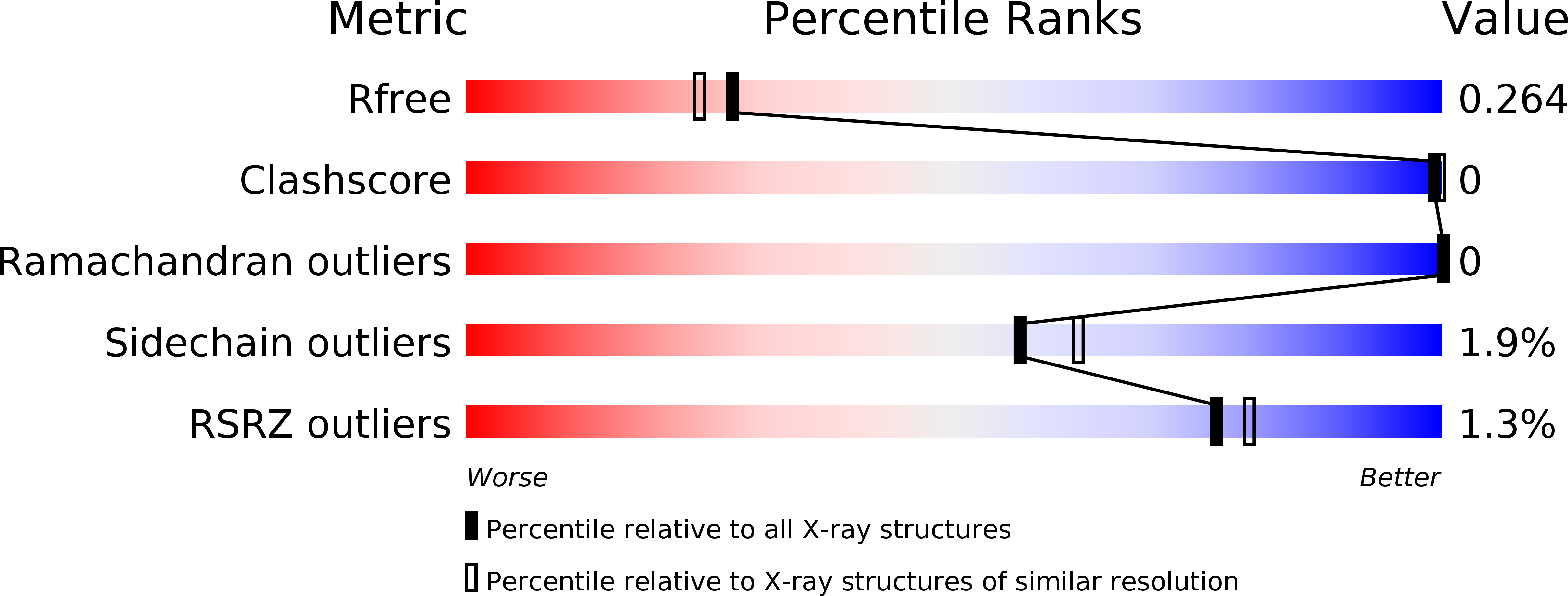
R-Value Free:
0.25
R-Value Work:
0.19
R-Value Observed:
0.19
Space Group:
P 1