5E1K image
Deposition Date 2015-09-29
Release Date 2015-11-25
Last Version Date 2024-10-16
Entry Detail
PDB ID:
5E1K
Title:
Selenomethionine Ca2+-Calmodulin from Paramecium tetraurelia SAD data
Biological Source:
Source Organism:
Host Organism:
Method Details:
Experimental Method:
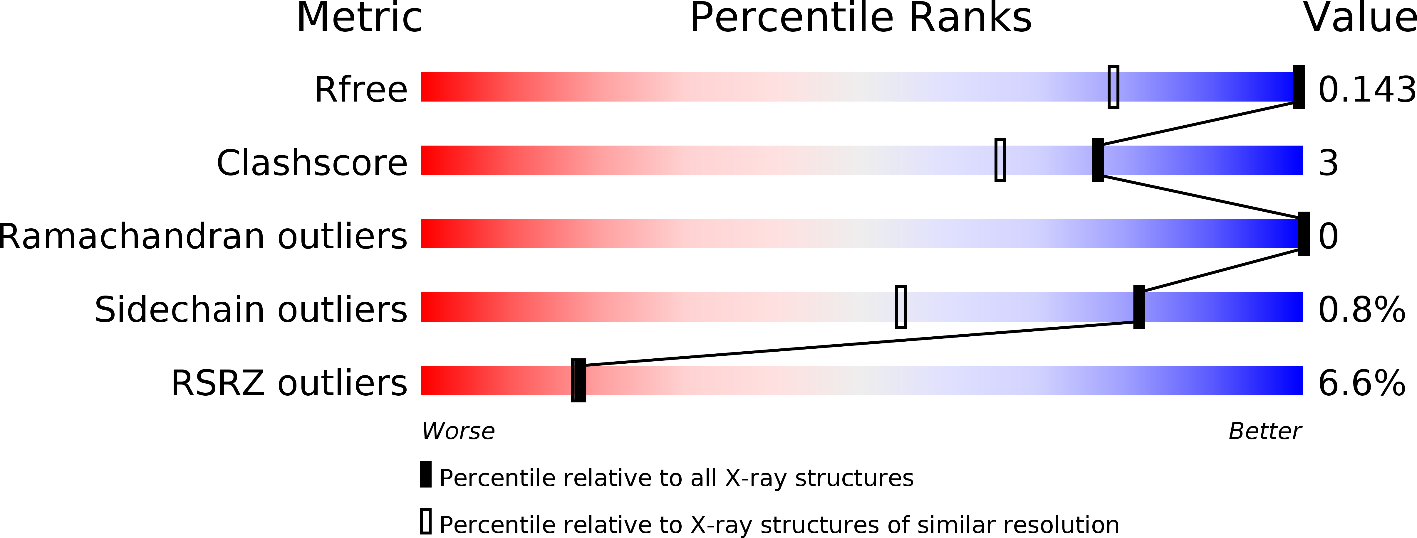
Resolution:
1.00 Å
R-Value Free:
0.16
R-Value Work:
0.14
R-Value Observed:
0.14
Space Group:
P 1
Macromolecular Entities
Polymer Type:polypeptide(L)
Molecule:Calmodulin
Gene (Uniprot):CAM
Chain IDs:A
Chain Length:148
Number of Molecules:1
Biological Source:Paramecium tetraurelia
Modified Residue
Compound ID Chain ID Parent Comp ID Details 2D Image
MSE A MET modified residue
Primary Citation
Atomic resolution experimental phase information reveals extensive disorder and bound 2-methyl-2,4-pentanediol in Ca(2+)-calmodulin.
Acta Crystallogr D Struct Biol 72 83 92 (2016)
PMID: 26894537 DOI: 10.1107/S2059798315021609

Abstact

Calmodulin (CaM) is the primary calcium signaling protein in eukaryotes and has been extensively studied using various biophysical techniques. Prior crystal structures have noted the presence of ambiguous electron density in both hydrophobic binding pockets of Ca(2+)-CaM, but no assignment of these features has been made. In addition, Ca(2+)-CaM samples many conformational substates in the crystal and accurately modeling the full range of this functionally important disorder is challenging. In order to characterize these features in a minimally biased manner, a 1.0 Å resolution single-wavelength anomalous diffraction data set was measured for selenomethionine-substituted Ca(2+)-CaM. Density-modified electron-density maps enabled the accurate assignment of Ca(2+)-CaM main-chain and side-chain disorder. These experimental maps also substantiate complex disorder models that were automatically built using low-contour features of model-phased electron density. Furthermore, experimental electron-density maps reveal that 2-methyl-2,4-pentanediol (MPD) is present in the C-terminal domain, mediates a lattice contact between N-terminal domains and may occupy the N-terminal binding pocket. The majority of the crystal structures of target-free Ca(2+)-CaM have been derived from crystals grown using MPD as a precipitant, and thus MPD is likely to be bound in functionally critical regions of Ca(2+)-CaM in most of these structures. The adventitious binding of MPD helps to explain differences between the Ca(2+)-CaM crystal and solution structures and is likely to favor more open conformations of the EF-hands in the crystal.

Legend

Protein

Chemical

Disease

Primary Citation of related structures